预约演示
更新于:2025-05-07

Carl Zeiss Meditec AG
更新于:2025-05-07
概览
标签
其他疾病
遗传病与畸形
眼部疾病
小分子化药
疾病领域得分
一眼洞穿机构专注的疾病领域

暂无数据
技术平台
公司药物应用最多的技术
靶点
公司最常开发的靶点
关联
靶点 |
作用机制 |
在研机构 |
原研机构 |
在研适应症 |
非在研适应症 |
最高研发阶段 |
首次获批国家/地区 |
首次获批日期 |

靶点 |
作用机制 |
在研机构 |
原研机构 |
在研适应症 |
非在研适应症 |
最高研发阶段 |
首次获批国家/地区 |
首次获批日期 |
CTRI/2024/07/070509
Post-Market Evaluation of Topography-Guided Treatment with the MEL Excimer Laser in Normal Virgin Eyes. - NIL
NCT06264232
A Multi-center, Prospective, Randomized, Controlled Clinical Trial to Demonstrate the Safety and Effectiveness of the Full Visual Range AT ELANA 841P Posterior Chamber Intraocular Lens for Correction of Aphakia Following Cataract Removal
NCT06428955
Prospective Multicenter Evaluation of the Visual Performance of a Non-constant Aberration Correcting Aspheric Monofocal Intraocular Lens (Precise Study)
100 项与 Carl Zeiss Meditec AG 相关的临床结果
登录后查看更多信息
登录后查看更多信息
2025-05-01Spectrochimica Acta Part A: Molecular and Biomolecular Spectroscopy
Enhancing surgical precision in squamous cell carcinoma of the head and neck: Hyperspectral imaging and artificial intelligence for improved margin assessment in an ex vivo setting
Article
作者: Alperovich, Anna ; Lehner, René ; Mostafa, Mayar ; Schwamborn, Carolin ; Zhang, Xiaohan ; Giannantonio, Tommaso ; Lingl, Julia ; Hoffmann, Thomas K ; Schuler, Patrick J ; Boehm, Felix
2025-02-01Journal of Cataract & Refractive Surgery
Prediction of visual outcomes using virtual implantation of a trifocal intraocular lens in presbyopic lens exchange patients
Article
作者: Schallhorn, Steven C ; Fernández, Joaquín ; Kaymak, Hakan ; Gerlach, Mario ; Kirchner, Friedrich O
2025-02-01Biomedical Optics Express
Development of a label-free, functional, molecular and structural imaging system combining optical coherence tomography and Raman spectroscopy for in vivo measurement of rat retina
Article
作者: Drexler, Wolfgang ; Shynkar, Vasyl ; Schmitt, Michael ; Sentosa, Ryan ; Salas, Matthias ; Schmoll, Tilman ; Baumann, Bernhard ; Unterhuber, Angelika ; Krestnikov, Igor ; Eibl, Matthias ; Merkle, Conrad W ; Kempe, Michael ; de Jong, Wim ; Amelink, Arjen ; Popp, Jürgen ; Leitgeb, Rainer A ; Andreana, Marco
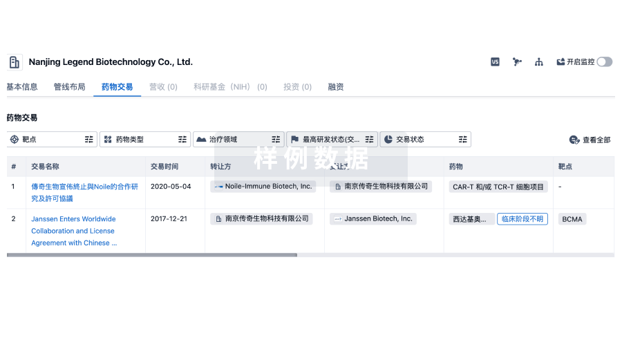
100 项与 Carl Zeiss Meditec AG 相关的药物交易
登录后查看更多信息
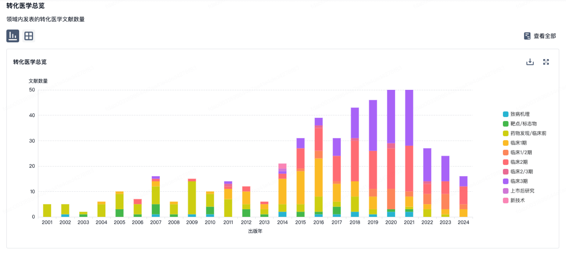
100 项与 Carl Zeiss Meditec AG 相关的转化医学
登录后查看更多信息
组织架构
使用我们的机构树数据加速您的研究。
登录
或

管线布局
2025年07月20日管线快照
管线布局中药物为当前组织机构及其子机构作为药物机构进行统计,早期临床1期并入临床1期,临床1/2期并入临床2期,临床2/3期并入临床3期
批准上市
2
登录后查看更多信息
当前项目
登录后查看更多信息
药物交易
使用我们的药物交易数据加速您的研究。
登录
或
转化医学
使用我们的转化医学数据加速您的研究。
登录
或
营收
使用 Synapse 探索超过 36 万个组织的财务状况。
登录
或
科研基金(NIH)
访问超过 200 万项资助和基金信息,以提升您的研究之旅。
登录
或
投资
深入了解从初创企业到成熟企业的最新公司投资动态。
登录
或
融资
发掘融资趋势以验证和推进您的投资机会。
登录
或
Eureka LS:
全新生物医药AI Agent 覆盖科研全链路,让突破性发现快人一步
立即开始免费试用!
智慧芽新药情报库是智慧芽专为生命科学人士构建的基于AI的创新药情报平台,助您全方位提升您的研发与决策效率。
立即开始数据试用!
智慧芽新药库数据也通过智慧芽数据服务平台,以API或者数据包形式对外开放,助您更加充分利用智慧芽新药情报信息。
生物序列数据库
生物药研发创新
免费使用
化学结构数据库
小分子化药研发创新
免费使用