预约演示
更新于:2025-08-14

Mie University
更新于:2025-08-14
概览
标签
肿瘤
消化系统疾病
皮肤和肌肉骨骼疾病
放射与诊断药物
小分子化药
TCR-T细胞疗法
疾病领域得分
一眼洞穿机构专注的疾病领域

技术平台
公司药物应用最多的技术
靶点
公司最常开发的靶点
关联
靶点 |
作用机制 |
在研机构 |
原研机构 |
在研适应症 |
非在研适应症 |
最高研发阶段 |
首次获批国家/地区 |
首次获批日期 |

靶点 |
作用机制 |
在研机构 |
原研机构 |
在研适应症 |
非在研适应症 |
最高研发阶段 |
首次获批国家/地区 |
首次获批日期 |
US20240181087
专利挖掘靶点 |
作用机制 |
在研机构 |
原研机构 |
在研适应症 |
非在研适应症 |
最高研发阶段 |
首次获批国家/地区 |
首次获批日期 |
JPRN-UMIN000052914
Effect of body position exchange on dyspnea associated with single-sided pleural effusion in cancer patients: A randomized, crossover, controlled trial - Effect of body position exchange on dyspnea associated with single-sided pleural effusion in cancer patients: A randomized, crossover, controlled trial
JPRN-UMIN000054691
Psychological Status of Patients Undergoing Cancer Genomic Profiling: Qualitative study - Psychological Status of Patients Undergoing Cancer Genomic Profiling: Qualitative study
JPRN-UMIN000054615
Oral care for people with dementia: a scoping review - Oral care for people with dementia: a scoping review
100 项与 Mie University 相关的临床结果
登录后查看更多信息
登录后查看更多信息
2025-12-01PARASITOLOGY INTERNATIONAL
Chemotaxis of Miamiensis avidus to hyaluronic acid, a component of fish surface mucus
Article
作者: Kitamura, Shin-Ichi ; Kim, Sang-Hee ; Isshiki, Tadashi
Miamiensis avidus is a marine parasitic ciliate belonging to the order Scuticociliatida, the members of which are the causal agents of scuticociliatosis. This ciliate has resulted in considerable economic losses to the aquaculture industry in the Republic of Korea and Japan. Nevertheless, the development of vaccines and therapeutics has proven to be challenging, and there are currently no reports of such products commercially available in Korea or Japan. Furthermore, the aetiology and pathogenesis of M. avidus infection in fish remain unknown, as do the underlying reasons for the initial infection. Our goal was to investigate the mechanism of infection by examining the chemotactic response of M. avidus to fish tissues. We found that M. avidus exhibited a high degree of chemotaxis towards the tissues, blood, and surface mucus of the olive flounder (Paralichthys olivaceus) specimens used in the experiment. We analyzed the components of olive flounder surface mucus to determine the degree of chemotaxis induced by each component. Miamiensis avidus exhibited pronounced chemosensitivity to hyaluronic acid, one of the constituents. Given the nature of the surface mucus, it is plausible to suggest that it acts as a potent attractant for initial parasitism by scuticociliates.
2025-12-01IBRO Neuroscience Reports
Effect of L-theanine on cerebellar granule cell migration related to cognitive disorders
Article
作者: Tomoko, Matsuda ; Umekawa, Hayato ; Ibrahim, Mai ; Nishio, Masahiro ; Makoto, Ozeki ; Aya, Abe ; Kenji, Kuriya
Introduction:
Cerebellar granule cell migration plays a crucial role in cerebellum development, and any abnormalities in CGC migration can lead to significant neurological disorders such as anxiety, a common psychological disorder that impacts a person's emotional, physical, and social health. L-theanine, an amino acid found in green tea, demonstrates neuroprotective properties and regulates the release of neurotransmitters by stimulating CGC migration. This study investigated the impact of L-theanine on CGC migration related to cognitive disorders.
Methods:
ddY male mice treated with a single oral dose of L-theanine at varying concentrations (10 mg/kg) were assessed for anxiety, learning, and memory using the maze test and the Morris Water Maze test, where the average completion time and escape time of the mice were considered indicators of cognitive performance. CGC microexplants were isolated from newly born C57BL/N6 mice and treated with a series of increasing concentrations of L-theanine. The migration distance of the CGC under the different L-theanine concentrations was assessed after 24, 48, and 72 h post-treatment using phase-contrast microscopy and image analysis software.
Results and conclusion:
Mice's anxiety symptoms improved based on their performance on the maze test after treatment with L-theanine at 5 mg/ml. However, L-theanine at 0.05 mg/ml enhanced learning and memory abilities. Compared to other concentrations, L-theanine at 1 µM yielded the longest migration distance for CGC in vitro. Therefore, L-Theanine may serve as a potential therapeutic agent in supporting cerebellar development and enhancing cognitive skills. Further investigation is required to fully elucidate the molecular mechanisms and therapeutic potential of L-theanine in neurodevelopmental disorders.
2025-10-01SURGERY
Prognosis of a deep excision margin within or beyond subcutaneous fat for invasive acral melanoma of the sole: A multi-institutional retrospective study
Article
作者: Yamazaki, Naoya ; Kaneko, Takahide ; Yamamoto, Yuki ; Manabe, Keiko ; Nagai, Makoto ; Inozume, Takashi ; Funakoshi, Takeru ; Hiura, Azusa ; Asagoe, Kenji ; Kiniwa, Yukiko ; Umeda, Yoshiyasu ; Matsushita, Shigeto ; Nakagawa, Masahiro ; Minami, Shoichiro ; Nakamura, Yasuhiro ; Nakano, Eiji ; Ogata, Dai ; Kuwatsuka, Yutaka ; Ishizuki, Shoichiro ; Kishi, Akiko ; Doi, Reiichi ; Yamamoto, Yosuke ; Takenouchi, Tatsuya ; Uhara, Hisashi ; Kadono, Takafumi ; Koizumi, Shigeru ; Hatta, Naohito ; Hoashi, Toshihiko ; Kokubu, Hiraku ; Takai, Toshihiro ; Arima, Masaru ; Asai, Jun ; Iwasawa, Utsugi ; Iino, Shiro ; Ito, Takamichi ; Uchi, Hiroshi ; Ishikawa, Masashi ; Maekawa, Takeo ; Haga, Takahiro ; Ito, Shusaku ; Kitagawa, Hiroshi ; Nakagawa, Tomoe ; Sato, Sayuri ; Yasuda, Masahito ; Nakama, Kenta ; Inafuku, Kazuhiro ; Miyagawa, Takuya ; Fukushima, Satoshi ; Ichigozaki, Yuki
BACKGROUND:
Preservation of plantar subcutaneous fat is crucial for cushioning in the surgical treatment of acral melanoma of the sole. However, no studies exist on the relationship between deep margins and prognosis. We aimed to retrospectively compare the prognoses of different deep margins (within or beyond the subcutaneous fat) in patients with invasive acral melanoma of the sole who underwent wide local excision.
METHODS:
In this multi-institutional retrospective study, survival was compared between 2 groups of patients: those with tumors excised within (S group) and those beyond the subcutaneous fat (D group).
RESULTS:
In total, 464 patients were included. Cox multivariable analyses showed that the depth of the deep excision margin was not associated with local recurrence-free survival, overall survival, or distant metastasis-free survival (hazard ratios of 1.20, P = .36; 1.10, P = .66; and 1.42, P = .05, respectively). However, excision beyond the subcutaneous fat was negatively associated with disease-free survival (hazard ratio 1.45, P = .02). After propensity score matching (both groups, n = 139), no significant differences were observed in survival outcomes between the S and D groups (5-year local recurrence-free survival: 72.8 vs 66.8%, P = .55; 5-year disease-free survival: 55.3 vs 43.7%, P = .24; 5-year overall survival: 76.2 vs 73.2%, P = .52; 5-year distant metastasis-free survival: 63.3 vs 54.1%, P = .13). Subgroup analysis of American Joint Committee on Cancer stages revealed no significant differences in survival outcomes between the 2 groups at any stage.
CONCLUSION:
Wide local excision beyond the subcutaneous fat was not associated with survival benefit of acral melanoma of the sole. Excision within the subcutaneous fat may represent the optimal deep margin.
100 项与 Mie University 相关的药物交易
登录后查看更多信息
100 项与 Mie University 相关的转化医学
登录后查看更多信息
组织架构
使用我们的机构树数据加速您的研究。
登录
或
管线布局
2025年08月24日管线快照
管线布局中药物为当前组织机构及其子机构作为药物机构进行统计,早期临床1期并入临床1期,临床1/2期并入临床2期,临床2/3期并入临床3期
药物发现
2
1
临床前
临床2期
1
6
其他
登录后查看更多信息
当前项目
登录后查看更多信息
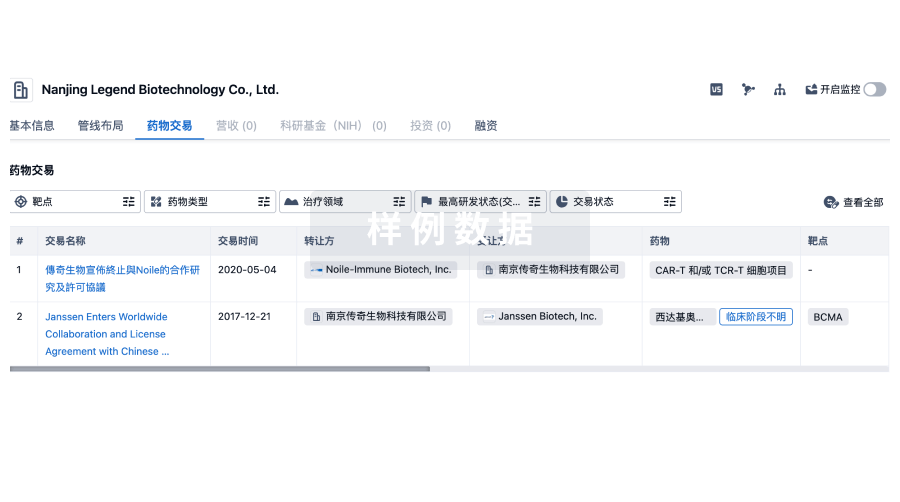
药物交易
使用我们的药物交易数据加速您的研究。
登录
或
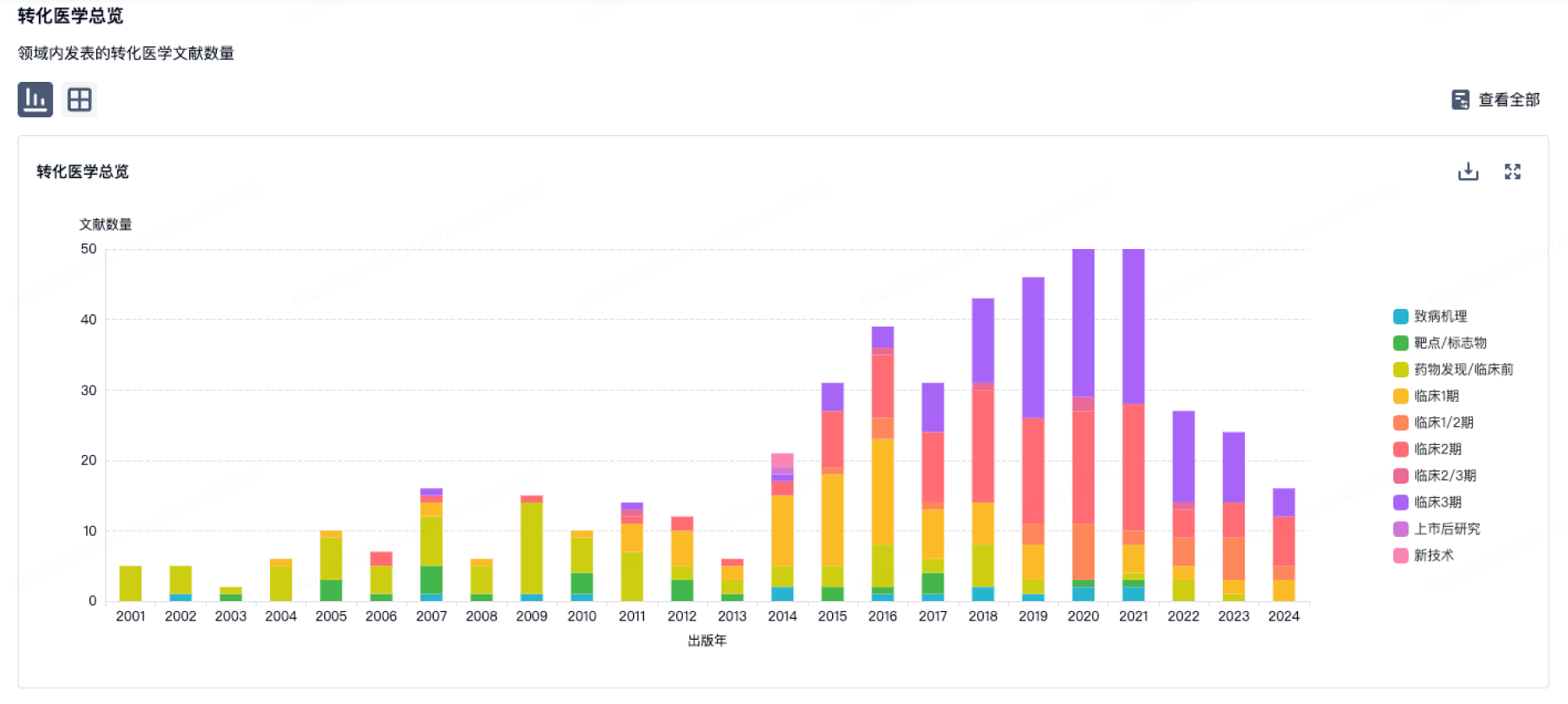
转化医学
使用我们的转化医学数据加速您的研究。
登录
或
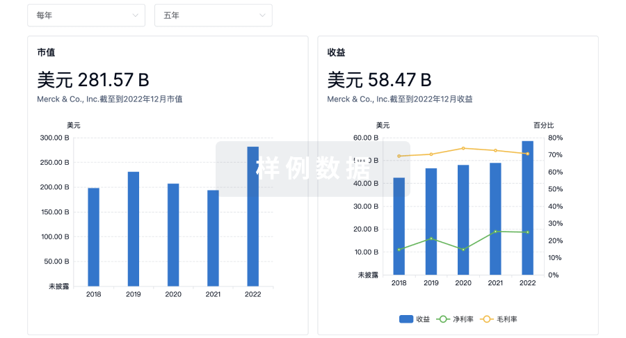
营收
使用 Synapse 探索超过 36 万个组织的财务状况。
登录
或
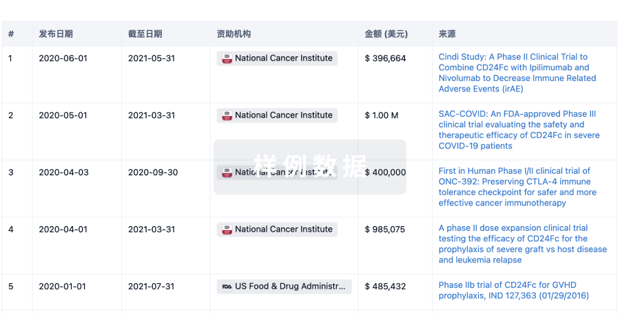
科研基金(NIH)
访问超过 200 万项资助和基金信息,以提升您的研究之旅。
登录
或
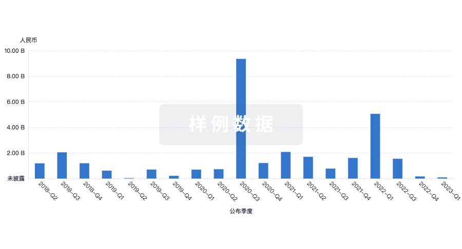
投资
深入了解从初创企业到成熟企业的最新公司投资动态。
登录
或
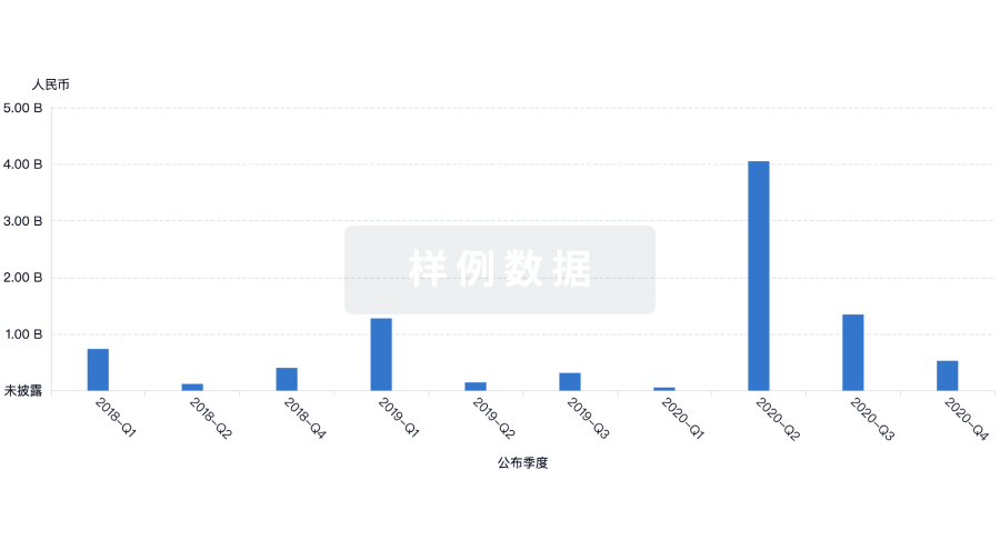
融资
发掘融资趋势以验证和推进您的投资机会。
登录
或
Eureka LS:
全新生物医药AI Agent 覆盖科研全链路,让突破性发现快人一步
立即开始免费试用!
智慧芽新药情报库是智慧芽专为生命科学人士构建的基于AI的创新药情报平台,助您全方位提升您的研发与决策效率。
立即开始数据试用!
智慧芽新药库数据也通过智慧芽数据服务平台,以API或者数据包形式对外开放,助您更加充分利用智慧芽新药情报信息。
生物序列数据库
生物药研发创新
免费使用
化学结构数据库
小分子化药研发创新
免费使用