预约演示
更新于:2025-05-07

Instituto Ramón y Cajal de Investigación Sanitaria
更新于:2025-05-07
概览
关联
靶点 |
作用机制 |
在研机构 |
原研机构 |
在研适应症 |
非在研适应症 |
最高研发阶段 |
首次获批国家/地区 |
首次获批日期 |

ISRCTN11587056
Surgery first and Invisalign. A comparative assessment of periodontal health and quality of life in postsurgical orthodontic treatment with Invisalign or traditional fixed appliances: a randomized controlled trial
100 项与 Instituto Ramón y Cajal de Investigación Sanitaria 相关的临床结果
登录后查看更多信息
登录后查看更多信息
2025-06-01Experimental Eye Research
P2 component latency of fVEP as a bioindicator for clinical and diagnostic use in visual pathologies
Article
作者: Liao, Fei ; Wang, Ting ; Liu, Haitao ; de la Villa, Pedro ; Germain, Francisco
2025-04-01Endoscopy
Single-use gastroscopes: evolution, revolution, or involution?
Article
作者: Rodriguez de Santiago, Enrique ; Pohl, Heiko
2025-04-01Actas Dermo-Sifiliográficas
[[Artículo traducido]]Revisión sistemática sobre suplementos dietéticos en la prevención y/o tratamiento de la queratosis actínica y el campo de cancerización
Review
作者: Longo, C ; Pellacani, G ; de Troya, M ; Peris, K ; de Galvez, M V ; Rodríguez-Luna, A ; Calzavara-Pinton, P ; Juarranz, Á ; Zamarrón, A ; Gilaberte, Y ; González, Salvador
100 项与 Instituto Ramón y Cajal de Investigación Sanitaria 相关的药物交易
登录后查看更多信息
100 项与 Instituto Ramón y Cajal de Investigación Sanitaria 相关的转化医学
登录后查看更多信息
组织架构
使用我们的机构树数据加速您的研究。
登录
或

管线布局
2025年07月21日管线快照
管线布局中药物为当前组织机构及其子机构作为药物机构进行统计,早期临床1期并入临床1期,临床1/2期并入临床2期,临床2/3期并入临床3期
其他
1
登录后查看更多信息
当前项目
登录后查看更多信息
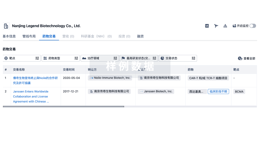
药物交易
使用我们的药物交易数据加速您的研究。
登录
或
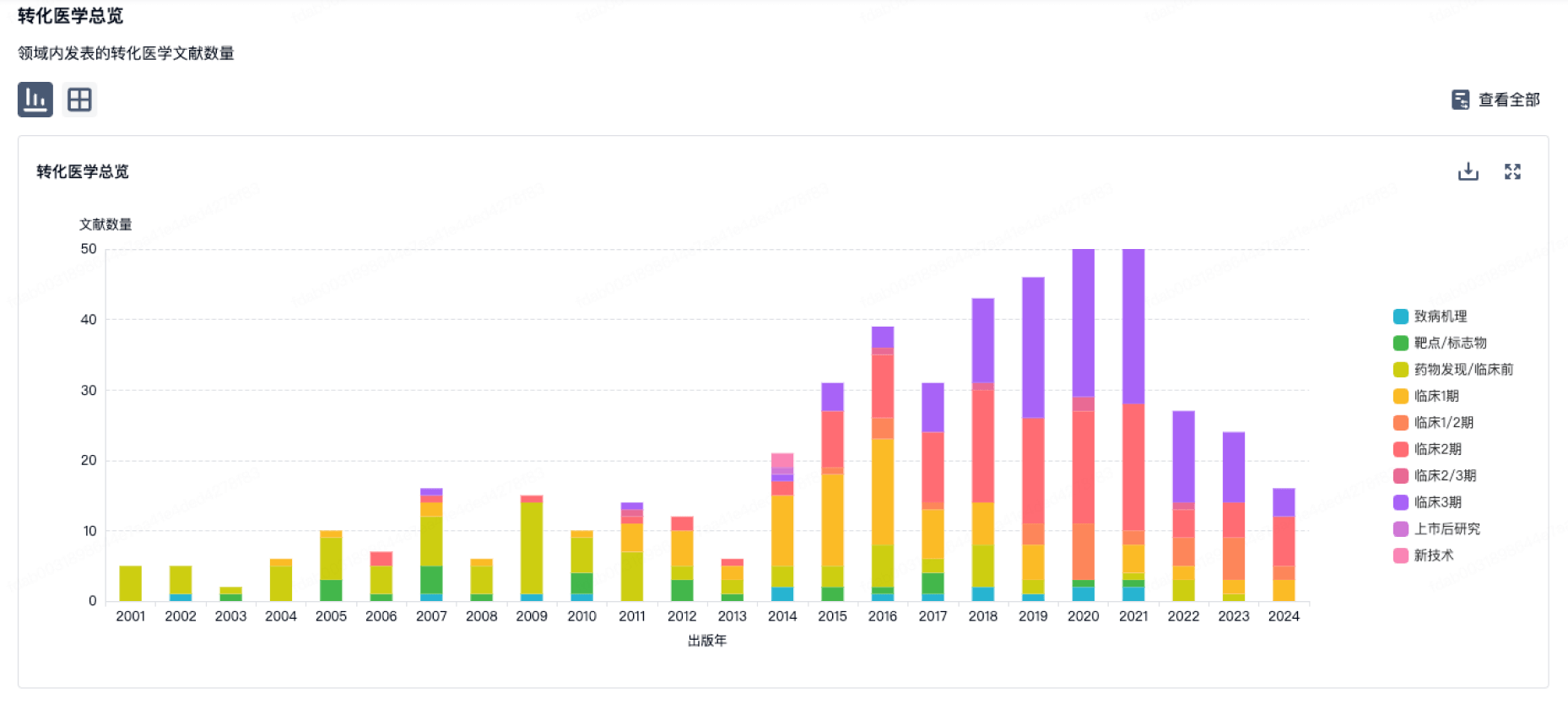
转化医学
使用我们的转化医学数据加速您的研究。
登录
或
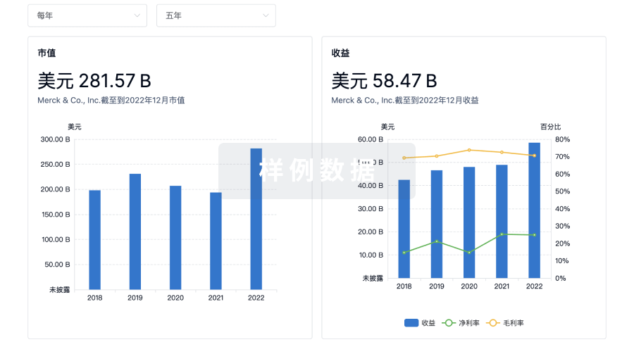
营收
使用 Synapse 探索超过 36 万个组织的财务状况。
登录
或
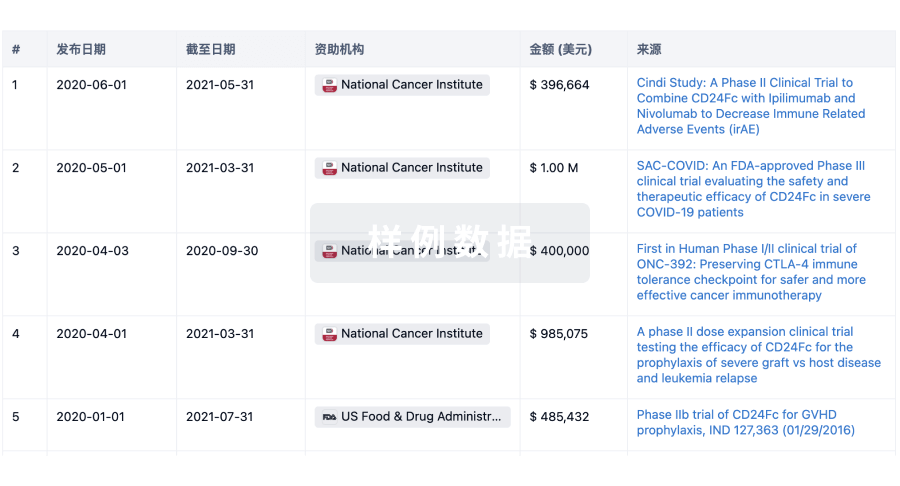
科研基金(NIH)
访问超过 200 万项资助和基金信息,以提升您的研究之旅。
登录
或
投资
深入了解从初创企业到成熟企业的最新公司投资动态。
登录
或
融资
发掘融资趋势以验证和推进您的投资机会。
登录
或
Eureka LS:
全新生物医药AI Agent 覆盖科研全链路,让突破性发现快人一步
立即开始免费试用!
智慧芽新药情报库是智慧芽专为生命科学人士构建的基于AI的创新药情报平台,助您全方位提升您的研发与决策效率。
立即开始数据试用!
智慧芽新药库数据也通过智慧芽数据服务平台,以API或者数据包形式对外开放,助您更加充分利用智慧芽新药情报信息。
生物序列数据库
生物药研发创新
免费使用
化学结构数据库
小分子化药研发创新
免费使用