预约演示
更新于:2025-09-09
Shanghai United Imaging Healthcare Co., Ltd.
更新于:2025-09-09
概览
标签
其他疾病
外泌体药物
疾病领域得分
一眼洞穿机构专注的疾病领域

暂无数据
技术平台
公司药物应用最多的技术
靶点
公司最常开发的靶点
暂无数据
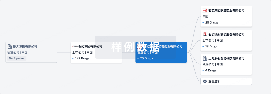
关联
靶点 |
作用机制 |
在研机构 |
原研机构 |
在研适应症 |
非在研适应症 |
最高研发阶段 |
首次获批国家/地区 |
首次获批日期 |

ChiCTR2500106556
Photon-counting energy spectrum CT clinical trial
ChiCTR2500098537
Clinical Investigation of deep learning reconstruction function in uCT 868
ChiCTR2500097616
A multicenter clinical study on the normal reference value of CT cardiac function in Chinese people
100 项与 上海联影医疗科技股份有限公司 相关的临床结果
登录后查看更多信息
登录后查看更多信息
2025-11-01APPLIED RADIATION AND ISOTOPES
Small field commissioning and verification for the uRT-TPOIS radiotherapy treatment planning system
Article
作者: Yang, Bo ; Liang, Yongguang ; Shi, Zhi ; Wen, Libin ; He, Shumeng ; Yu, Lang ; Liu, Yanfang ; Qiu, Jie ; Ye, Shaoqiang ; Mo, Zijie ; Yang, Jingru
PURPOSE:
Stereotactic radiosurgery (SRS) and stereotactic body radiation therapy (SBRT) plans involve high doses and small fields. Standard commissioning procedures typically do not prioritize small fields, which is why the commission parameters of small field models require a comprehensive and precise verification process to ensure accurate dose calculation for SRS/SBRT plans.
METHODS:
The study employed various methods, including dose validation tests in a water tank with both regular and irregular small fields. The StereoPHAN and SRS MapCHECK were utilized to assess the modelling accuracy of collapse cone convolution (CC) and Monte Carlo (MC) algorithms for both point and planar doses of the small field plans. Additionally, pre-treatment approval of SRS/SBRT plans based on Electronic Portal Imaging Device (EPID) equipped on the machine was conducted. To enhance realism, 19 clinical SRS/SBRT plans were measured and analyzed for their point and planar doses. In addition, a special 27-target plan was measured using 'gold standard' film and compared with the calculations based on parameters from the small field commissioning procedures.
RESULTS:
The validation tests conducted in the water tank proved to be reliable and accurate for both regular and irregular small fields. The results indicated that the average error of point dose decreased from 2.29 % with parameters based on standard commissioning procedures to 0.64 % with parameters based on small field commissioning procedures. Furthermore, planar doses acquisition included high-dose and non-high-dose planes, resulting in an increase in the average gamma passing rate from 93.15 % to 99.90 %. The gamma passing rates of EPID pre-treatment verifications exceeded 99 % at a criteria of 2mm2 %. These findings demonstrate the effectiveness of small field commissioning procedures in improving the accuracy of dose calculations for SRS/SBRT plans.
CONCLUSION:
This study marks the first time that small field commissions and verifications of CC and MC algorithms in uRT-TPOIS, along with uRT-linac 506c, were conducted. The reliability and accuracy of the calculation algorithms for small field commissions were verified through the comparisons between measurements and calculations. The results showed that both CC and MC algorithms' small field parameters met the clinical requirements with minimal deviations (lower than 2 %) between measurements and calculations. Additionally, EPID was found to be an effective quality assurance tool for SRS/SBRT plans.
2025-10-01EUROPEAN JOURNAL OF RADIOLOGY
Optimized DWI-based virtual MR elastography for diagnosis and therapeutic monitoring of focal liver lesions
Article
作者: Huang, Xutong ; Huang, Lingying ; Yuan, Jie ; Tan, Wenli ; Ma, Wenxin ; Qin, Zhiwei ; Lu, Shixian ; Cui, Yu
OBJECTIVES:
This study aimed to develop and evaluate an optimized DWI-based virtual MR elastography (vMRE) technique for diagnosing focal liver lesions (FLLs) and monitoring therapeutic responses in malignant tumors, comparing its performance with traditional MRE.
MATERIALS AND METHODS:
This retrospective analysis of 142 patients with FLLs using six b-value DWI (150-1500 s/mm2) to calculate eight apparent diffusion coefficient (ADC) combinations. Linear regression analysis was performed to evaluate the correlation between ADC and traditional MRE, and a vMRE formula was constructed. Diagnostic performance of vMRE and MRE was compared for FLLs. Prognostic capabilities of both techniques were assessed in malignant tumors using paired t-tests across three response groups at two time points.
RESULTS:
Significant negative correlations were found between ADC values and MRE stiffness (all P < 0.001), and strongest at b = 200/1500 s/mm2 (rho = - 0.785). vMRE and MRE effectively differentiated benign/malignant lesions (AUCs: 0.957 vs. 0.944, P = 0.440), with vMRE showing higher specificity (95.1 % vs. 85.4 %) and MRE better sensitivity (89.2 % vs. 86.3 %). Longitudinal analysis revealed MRE detected earlier stiffness reductions in partial responders (-31.0 %, P = 0.01), while vMRE showed a non-significant decrease (-12.5 %, P > 0.05). Both modalities identified significant stiffness escalation in progressors (vMRE + 40.1 %, MRE + 43.7 %, both P < 0.01) but not in stable disease (all P > 0.05).
CONCLUSIONS:
Optimized DWI-based vMRE effectively differentiates FLLs, offering high specificity for diagnosis and comparable prognostic capabilities to MRE in monitoring malignant tumor responses.
2025-10-01Journal of Plastic Reconstructive and Aesthetic Surgery
Magnetic resonance lymphangiography: Correlation with indocyanine green lymphangiography in lower limb lymphedema
Article
作者: Zhang, Minge ; Liu, Chong ; Wang, Jingjing ; Chen, Yan ; Yi, Liqi ; Tang, Runyu ; Huang, Jinbiao ; Yang, Hai
BACKGROUND:
Indocyanine green (ICG) lymphography is considered the gold standard method for real-time imaging of the lymphatic vessels. Despite extensive literature on the effectiveness of different imaging modalities, research comparing the reliability of ICG lymphography and magnetic resonance lymphangiography (MRL) is limited. We retrospectively analyzed and compared the capability of MRL and ICG lymphangiography to detect lower limb lymphoedema abnormalities.
METHODS:
This monocentric cohort study included patients who had examinations and surgeries between September 2021 and December 2023. The assessment comprised scoring the severity levels of the drainage pattern, lymphatic vessel conspicuity, anatomic level of lymphatic vessel enhancement, contrast medium deposition in the foot, and the anatomic coverage of dermal backflow. Sensitivity, specificity, positive predictive value (PPV), and negative predictive value (NPV) of MRL and ICG lymphangiography were calculated.
RESULTS:
A total of 89 patients with clinically proven moderate to severe lymphoedema (stages II-III) affecting 92 lower limbs were enrolled. Good to excellent agreement was determined between MRL and ICG lymphography in drainage pattern (k=0.710), contrast medium deposition in the foot (k=0.933), and anatomic coverage of dermal backflow (k=0.653). Anatomical level of lymphatic vessel enhancement was in moderate correlation (k=0.524), and the visualization of lymphatic vessels was correlated (k=0.330) between the two modalities. MRL demonstrated lower NPV (50% vs 100%) in lymphatic vessel conspicuity but better sensitivity (100% vs 98%) and NPV (100% vs 97.6%) of contrast medium deposition in the foot as compared to ICG lymphography.
CONCLUSIONS:
MRL demonstrated better performance on the lymphatic vessel conspicuity when dermal backflow is obvious. For normal lymphatic vessels, ICG lymphography provided a better description.
100 项与 上海联影医疗科技股份有限公司 相关的药物交易
登录后查看更多信息
100 项与 上海联影医疗科技股份有限公司 相关的转化医学
登录后查看更多信息
组织架构
使用我们的机构树数据加速您的研究。
登录
或
管线布局
2025年09月10日管线快照
管线布局中药物为当前组织机构及其子机构作为药物机构进行统计,早期临床1期并入临床1期,临床1/2期并入临床2期,临床2/3期并入临床3期
临床前
1
登录后查看更多信息
当前项目
登录后查看更多信息
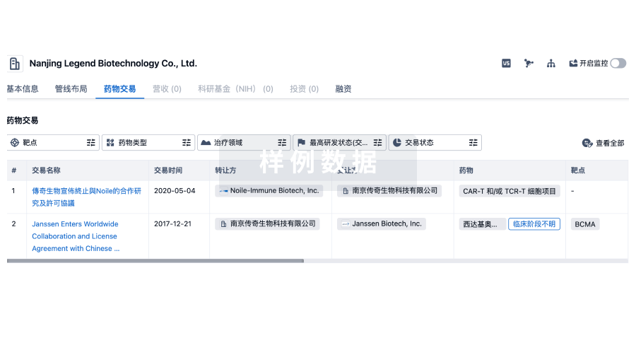
药物交易
使用我们的药物交易数据加速您的研究。
登录
或
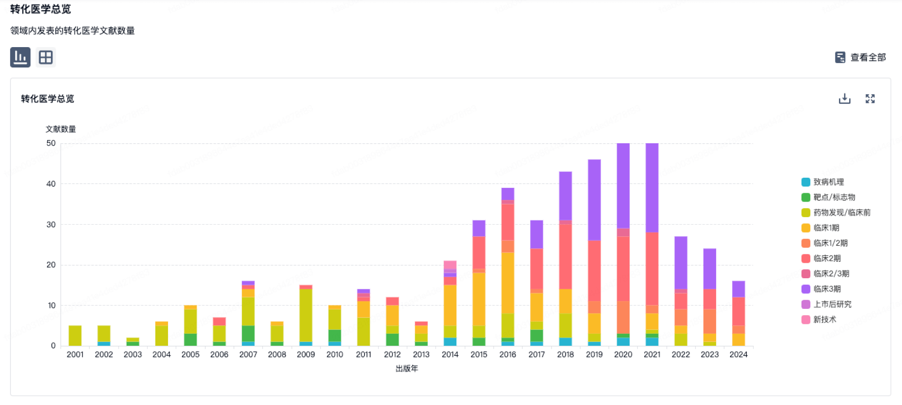
转化医学
使用我们的转化医学数据加速您的研究。
登录
或
营收
使用 Synapse 探索超过 36 万个组织的财务状况。
登录
或
科研基金(NIH)
访问超过 200 万项资助和基金信息,以提升您的研究之旅。
登录
或
投资
深入了解从初创企业到成熟企业的最新公司投资动态。
登录
或
融资
发掘融资趋势以验证和推进您的投资机会。
登录
或
Eureka LS:
全新生物医药AI Agent 覆盖科研全链路,让突破性发现快人一步
立即开始免费试用!
智慧芽新药情报库是智慧芽专为生命科学人士构建的基于AI的创新药情报平台,助您全方位提升您的研发与决策效率。
立即开始数据试用!
智慧芽新药库数据也通过智慧芽数据服务平台,以API或者数据包形式对外开放,助您更加充分利用智慧芽新药情报信息。
生物序列数据库
生物药研发创新
免费使用
化学结构数据库
小分子化药研发创新
免费使用