预约演示
更新于:2025-09-09
Wannan Medical College
更新于:2025-09-09
概览
标签
肿瘤
消化系统疾病
皮肤和肌肉骨骼疾病
小分子化药
化学药
合成多肽
疾病领域得分
一眼洞穿机构专注的疾病领域

技术平台
公司药物应用最多的技术
靶点
公司最常开发的靶点
关联
靶点 |
作用机制 |
在研机构 |
原研机构 |
在研适应症 |
非在研适应症 |
最高研发阶段 |
首次获批国家/地区 |
首次获批日期 |
靶点 |
作用机制 |
在研机构 |
原研机构 |
在研适应症 |
非在研适应症 |
最高研发阶段 |
首次获批国家/地区 |
首次获批日期 |
靶点 |
作用机制 |
在研机构 |
原研机构 |
在研适应症 |
非在研适应症 |
最高研发阶段 |
首次获批国家/地区 |
首次获批日期 |
NCT07127484
Efficacy and Safety of Hemoadsorption for Severe Ischemic Stroke
NCT07150624
Polyene Phosphatidylcholine Injection for the Treatment of Perioperative Liver Injury With Laparoscopic Hemihepatectomy in Hepatocellular Carcinoma: a Multicenter Randomized Controlled Study
ChiCTR2500107853
Exploring the pathogenesis of pediatric intussusception based on gut microbiota comparison: A Multicenter Retrospective and Prospective Cohort Study
100 项与 皖南医学院 相关的临床结果
登录后查看更多信息
登录后查看更多信息
2026-02-01BIOMATERIALS
Hydroxyl-augmented gold nanorods via lactone ring-opening alleviate cisplatin nephrotoxicity through Nrf2 activation
Article
作者: Cai, Yueming ; Fang, Chunyan ; Cai, Ying ; Lu, Yong ; Wang, Xiaoyan ; He, Cui
Cisplatin-induced acute kidney injury (AKI) epitomizes a critical barrier and paradox in cancers, where dose-limiting nephrotoxicity propelled by oxidative cascades lacks targeted interventions. This pressing clinical dilemma underscores the paramount need for kidney injury mitigation, while existing therapeutic strategies fail to achieve it due to inadequate efficacy and specificity. Here, we introduce a lactone ring-opening chemistry approach to amplify hydroxyl-driven redox modulation on gold nanorods (Au-M NRs), addressing the unmet need for precision antioxidant delivery. Specifically, the sequential functionalization of gold nanorods with thiolated polyethylene glycol amine (HS-PEG2000-NH2) and 5,8-dihydroxypsoralen (5,8-DHP) triggered a structural transformation, cleaving the six-membered lactone ring of 5,8-DHP and generating an additional phenolic hydroxyl group. In cisplatin-challenged HK-2 cells, hydroxyl-enriched Au-M markedly attenuated reactive oxygen species (ROS) level and activated Keap1/Nrf2 pathway, evidenced by upregulated SOD and antioxidant Nqo1 expression alongside suppressed MDA and prooxidant Nox2 levels. SiNrf2 transfection severely abrogated the cytoprotective effects of Au-M, while Nrf2 overexpression synergistically enhanced the anti-apoptotic efficacy of Au-M in renal tubular epithelial cells. Mirroring cellular outcomes, the Au-M nanosystem elicited potent renoprotection via Nrf2-orchestrated transcriptional reprogramming in vivo, concurrently preserving the integrity and safety of healthy tissues. Critically, Au-M NRs conferred sustained protection in chronic exposure models, exhibited biocompatibility upon repeated dosing, and accelerated renal recovery, affirming their long-term therapeutic viability. This lactone-to-phenol conversion strategy pioneers a topology-guided paradigm for redox homeostasis regulation beyond AKI, establishing a transformative platform to harness natural product derivatization in combating organ-specific oxidative injury.
2025-12-31REDOX REPORT
MS-275 facilitates osseointegration in osteoporotic rats by mitigating oxidative stress via activation of the miR-200a/Keap1/Nrf2 signaling pathway
Article
作者: Yan, Junjie ; Chen, Yuhu ; Guan, Wengang ; Chen, Bin ; Jiang, Wenkai ; Gu, Qinsong ; Yang, Min ; Zhou, Zhi ; Li, Jianqiao
OBJECTIVES:
Osteoporosis, a prevalent metabolic bone disease affecting millions worldwide. Although MS-275 has been reported to inhibit oxidative stress, its ability to protect osteoblasts from oxidative stress damage has yet to be clarified. This study investigated whether MS-275 can inhibit oxidative stress and promote osteogenesis by activating the miRNA-200a/Keap1/Nrf2 signaling pathway.
METHODS:
In vitro, MC3T3-E1 cells underwent induction with carbonyl cyanide 3-chlorophenylhydrazone, leading to the establishment of an oxidative stress model, investigating the underlying mechanism. In vivo, using a rat model of ovariectomized osteoporosis, evaluating the effects of MS-275.
RESULTS:
In vitro, MS-275 treatment of oxidation-induced MC3T3-E1 cells resulted in up-regulation of osteoblast protein, increased expression of miRNA-200a, increased binding of miRNA-200a to Keap1 mRNA, decreased expression of Keap1 protein, and dissociation of Nrf2 from Keap1. The expressions of total Nrf2, nuclear Nrf2 and HO-1 were increased, mitochondrial function was enhanced, and oxidative damage was reduced. However, these effects were reversed after interference with miRNA-200a. In vivo,MS-275 effectively enhanced the microstructural features of distal femoral trabecular bone, increased the mineralization capacity of osteoblasts, and promoted bone formation.
DISCUSSION:
MS-275 can reverse oxidative stress-induced cell damage, promote bone healing, and improve osteoporosis by activating the miRNA-200a/Keap1/Nrf2 pathway.
2025-12-31RENAL FAILURE
Fecal microbiota transplantation modulates myeloid-derived suppressor cells and attenuates renal fibrosis in a murine model
Article
作者: Chen, Yuye ; Cao, Yuhan ; Xiao, Zihao ; Wang, Yajie ; Fu, Cong ; Shi, Yuanhui
BACKGROUND:
Renal fibrosis is a hallmark of progressive chronic kidney disease (CKD), with emerging evidence linking gut microbiota dysbiosis to disease progression. Myeloid-derived suppressor cells (MDSCs) have demonstrated renoprotective effects, yet the impact of fecal microbiota transplantation (FMT) on MDSC-mediated modulation of renal fibrosis remains unclear.
METHODS:
C57BL/6J mice underwent unilateral ureteral obstruction (UUO) to induce renal fibrosis, followed by FMT administration via gavage. Flow cytometry was used to quantify granulocytic (G-MDSCs) and monocytic (M-MDSCs) MDSC populations in peripheral blood, kidney, and spleen. To elucidate the role of MDSCs in FMT-mediated effects, MDSCs were depleted or adoptively transferred in vivo. Renal fibrosis severity and inflammatory cytokine expression were subsequently analyzed.
RESULTS:
FMT altered MDSC distribution, increasing M-MDSC accumulation in the blood and kidney. This was associated with downregulation of proinflammatory cytokines and attenuation of renal fibrosis. Adoptive MDSC transfer similarly produced anti-inflammatory and antifibrotic effects, reinforcing their therapeutic role in FMT-mediated renal protection.
CONCLUSIONS:
FMT enhances M-MDSC-mediated immunomodulation, reducing inflammation and renal fibrosis in UUO-induced CKD. These findings suggest a potential therapeutic strategy targeting the gut-kidney axis in CKD management.
100 项与 皖南医学院 相关的药物交易
登录后查看更多信息
100 项与 皖南医学院 相关的转化医学
登录后查看更多信息
组织架构
使用我们的机构树数据加速您的研究。
登录
或
管线布局
2025年11月01日管线快照
管线布局中药物为当前组织机构及其子机构作为药物机构进行统计,早期临床1期并入临床1期,临床1/2期并入临床2期,临床2/3期并入临床3期
临床前
7
1
早期临床1期
其他
1
登录后查看更多信息
当前项目
登录后查看更多信息
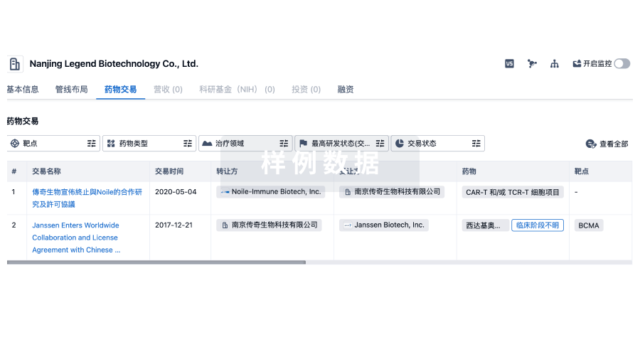
药物交易
使用我们的药物交易数据加速您的研究。
登录
或
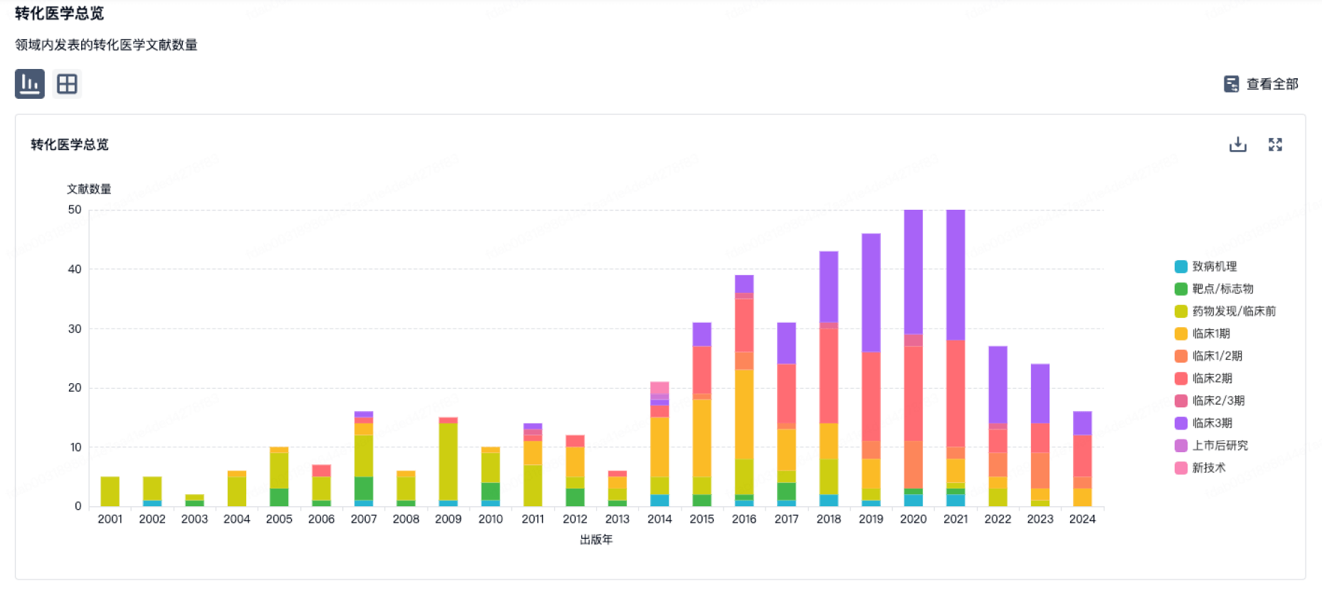
转化医学
使用我们的转化医学数据加速您的研究。
登录
或
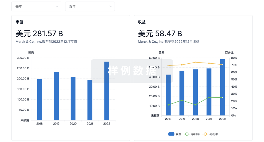
营收
使用 Synapse 探索超过 36 万个组织的财务状况。
登录
或
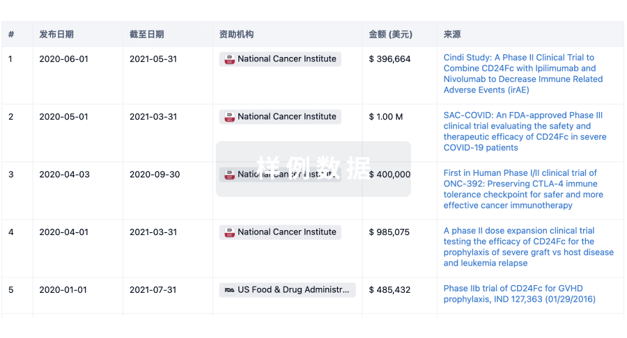
科研基金(NIH)
访问超过 200 万项资助和基金信息,以提升您的研究之旅。
登录
或
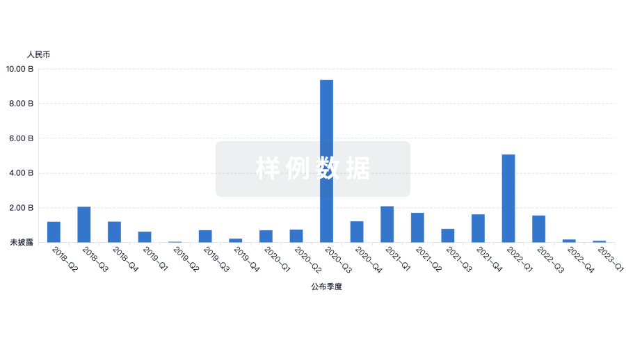
投资
深入了解从初创企业到成熟企业的最新公司投资动态。
登录
或
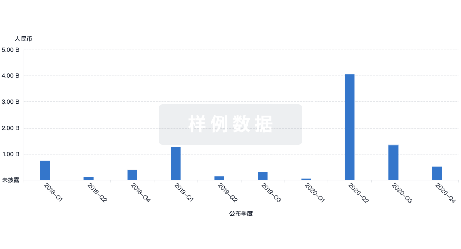
融资
发掘融资趋势以验证和推进您的投资机会。
登录
或
生物医药百科问答
全新生物医药AI Agent 覆盖科研全链路,让突破性发现快人一步
立即开始免费试用!
智慧芽新药情报库是智慧芽专为生命科学人士构建的基于AI的创新药情报平台,助您全方位提升您的研发与决策效率。
立即开始数据试用!
智慧芽新药库数据也通过智慧芽数据服务平台,以API或者数据包形式对外开放,助您更加充分利用智慧芽新药情报信息。
生物序列数据库
生物药研发创新
免费使用
化学结构数据库
小分子化药研发创新
免费使用