预约演示
更新于:2025-03-08
Siberian State Medical University
更新于:2025-03-08
概览
标签
肿瘤
泌尿生殖系统疾病
多肽偶联核素
诊断用放射药物
疾病领域得分
一眼洞穿机构专注的疾病领域

技术平台
公司药物应用最多的技术
靶点
公司最常开发的靶点
关联
靶点 |
作用机制 |
在研机构 |
原研机构 |
在研适应症 |
非在研适应症 |
最高研发阶段 |
首次获批国家/地区 |
首次获批日期 |

NCT04476992
A Safety Study on the Use of Intermittent Versus Continuous Inhalation of NO in Spontaneous Breathing COVID-19 Patients
NCT03855046
A Comparative Efficacy and Safety Study of Lateral Subcutaneous Sphincterotomy and Botulinum Toxin Type A in the Treatment of Chronic Anal Fissure

ISRCTN10577372
Dose-finding and pharmacokinetic studies of praziquantel in patients infected with Opisthorchis felineus
100 项与 Siberian State Medical University 相关的临床结果
登录后查看更多信息
登录后查看更多信息
2025-12-01HISTOCHEMISTRY AND CELL BIOLOGY
A hypothesis of mesenchymal stem cell osteogenic differentiation mediated by chelidonic acid through the calcium import: original research and computer simulation
Article
作者: Avdeeva, Elena ; Shupletsova, Valeria ; Litvinova, Larisa ; Belousov, Mikhail ; Gorokhova, Anna ; Bariev, Usman ; Porokhova, Ekaterina ; Nasibov, Temur ; Khlusov, Igor ; Yurova, Kristina
Chelidonic acid (ChA) is small molecule capable of inducing the differentiation of mesenchymal stem cells (MSCs) into osteoblasts and the formation of mineralized bone matrix (MBM) both in vitro and in vivo. However, the molecular mechanisms underlying these effects are unknown. Therefore, in silico modelling of potential molecular targets of ChA was performed. ChA was isolated from Saussurea controversa. The ability of ChA to induce in vitro differentiation MSCs into osteoblasts synthesizing MBM was detected using alizarin red staining. ChA osteogenic activity was studied in mice by in situ test of ectopic osteogenesis, using the subcutaneous implantation of syngeneic bone marrow on the calcium phosphate coated titanium plates. DIGEP-Pred web service was used to simulate in silico the effect of ChA on gene expression, and overrepresentation analysis to search for common ontologies and pathways. ChA linearly increased the number of single (R2 = 0.92, p = 0.039) and the total areas of MBM sites (R2 = 0.96, p = 0.019) in a 21-day MSC culture. Oral administration of ChA led to two to three times improved bone and bone marrow formation in situ. In silico modelling identified 306 genes (including 7 calcium import genes) and 9 signalling pathways potentially involved in ChA osteogenic effect and calcium metabolism in MSCs. In silico analysis revealed a list of key signalling pathways and genes for calcium influx into MSCs and their differentiation into osteoblasts as the first target candidates for studying real gene expression and molecular mechanisms of the ChA osteogenic effects.
2025-03-06Zhurnal nevrologii i psikhiatrii imeni S.S. Korsakova
A variant of p.Arg1623Gln of the DYNC1H1 gene in a patient with corpus callosum agenesis, polydactyly, mental development disorder, and neuromuscular system disorders
Article
作者: Fonova, E.A. ; Zuev, A.S. ; Sivokha, V.M. ; Minaycheva, L.I. ; Podus, A.A. ; Lebedev, I.N. ; Demeneva, V.V. ; Seitova, G.N. ; Kraeva, L.S. ; Zarubin, A.A.
DYNC1H1 encodes the heavy chain of dynein 1, a protein that plays a critical role in intracellular transport and is also involved in neurogenesis, bipolar spindle apparatus formation, and interaction with certain regulatory proteins. Many variants of the gene are described in various neuromuscular, psychoneurological, congenital abnormalities, and malignancies. In this clinical case, the correlation of clinical manifestations with molecular genetic changes of the DYNC1H1 gene was evaluated. A variant was identified and presented de novo in the linker domain of the protein in a patient with abnormalities of brain development (pachygyria of the temporal and frontal lobes, polymicrogyria of the occipital lobes, cerebellar agenesis), polydactylitis, mental development disorder, and involvement of the neuromuscular system, as well as congenital cataracts. In this case, a new feature is described — polydactyly — not previously described in the variant c.4868G>A (p.Arg1623Gln), which expands the range of clinical manifestations and can contribute to the understanding of the mechanisms of phenotypic heterogeneity, as well as the development of optimal diagnostic algorithms.
2025-02-11Zhurnal nevrologii i psikhiatrii imeni S.S. Korsakova
Indicators of the background spectral power of the EEG in patients with alcohol dependence complicated by exogenous organic brain disease
Article
作者: Aksenov, M.M. ; Perchatkina, O.E. ; Bokhan, N.A. ; Galkin, S.A. ; Mandel, A.I.
Objective. To conduct a comparative analysis of EEG spectral power indicators in patients with alcoholism with- or without exogenous organic brain disease. Material and methods. An analysis of 119 EEG of patients with alcohol dependence was carried out. The presence of exogenous organic brain disease in patients was assessed retrospectively (through anamnesis). The EEG signals received from 16 electrodes were first divided into 3 frequency spectra (i.e. theta, alpha and beta), then the values of absolute spectral power were calculated, and then they were reordered, combined and topographically transferred to a spectral map. Data analysis was carried out in 5 different ways: electrode analysis, analysis based on cortical regions, lateralization, antero-posterior locus and throughout the whole cortex. Results. In the group of alcohol-dependent patients with comorbid exogenous organic brain damage, significantly (p<0.05) lower values of the spectral power of alpha activity on the Fp2, F3, F4, C3 and C4 electrodes as well as higher values of the spectral power of beta activity on the F8, T5 and T6 electrodes compared with the group of patients with «pure» alcohol dependence were observed. The intergroup data analysis, taking into account the topographic distribution, revealed significant differences in the spectral power of alpha activity in the prefrontal (F=4.201; p=0.043), frontal (F=3.951; p=0.049), central (F=4.033; p=0.047) and parietal (F=3.926; p=0.049) regions, as well as beta activity in the temporal region (F=5.656; p=0.019) of the cerebral cortex. Compared with patients suffering only from alcohol dependence, the group of patients with alcohol dependence and exogenous organic brain damage had significantly lower spectral power of alpha activity on the left (F=5.329; p=0.019) and on the right (F=5.191; p=0.027), as well as in the anterior locus (F=3.975; p=0.049). Conclusion. The results clearly indicate that the presence of exogenous organic brain damage in patients with alcohol dependence is accompanied by a change in the spectral power of the EEG mainly in the alpha frequency range.
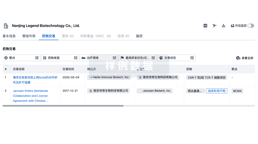
100 项与 Siberian State Medical University 相关的药物交易
登录后查看更多信息
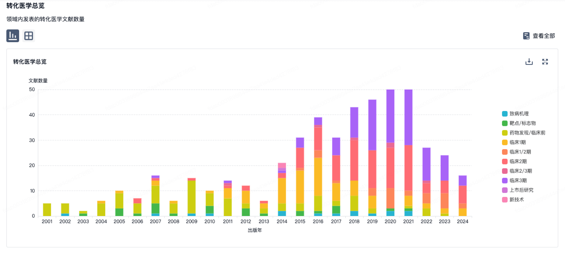
100 项与 Siberian State Medical University 相关的转化医学
登录后查看更多信息
组织架构
使用我们的机构树数据加速您的研究。
登录
或
管线布局
2025年04月09日管线快照
管线布局中药物为当前组织机构及其子机构作为药物机构进行统计,早期临床1期并入临床1期,临床1/2期并入临床2期,临床2/3期并入临床3期
临床前
1
1
其他
登录后查看更多信息
当前项目
登录后查看更多信息
药物交易
使用我们的药物交易数据加速您的研究。
登录
或
转化医学
使用我们的转化医学数据加速您的研究。
登录
或
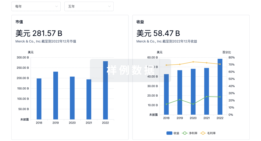
营收
使用 Synapse 探索超过 36 万个组织的财务状况。
登录
或
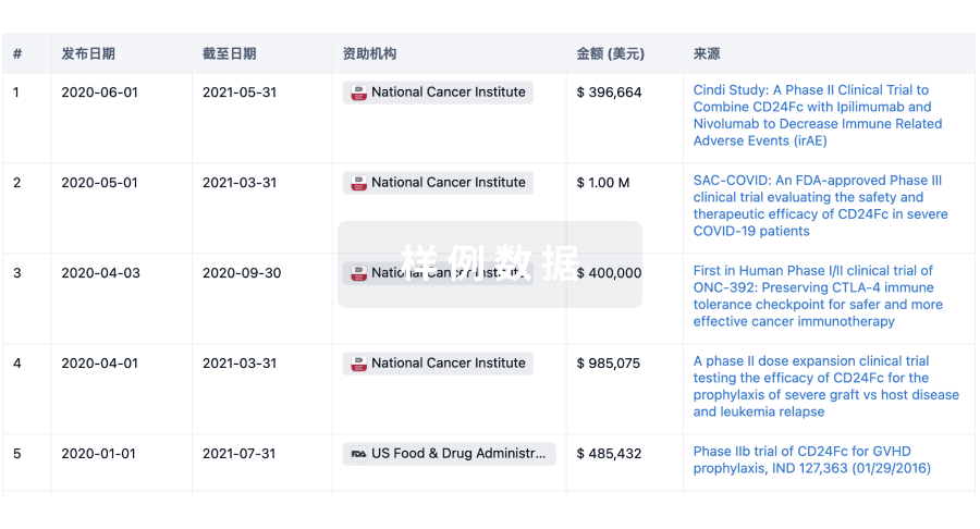
科研基金(NIH)
访问超过 200 万项资助和基金信息,以提升您的研究之旅。
登录
或
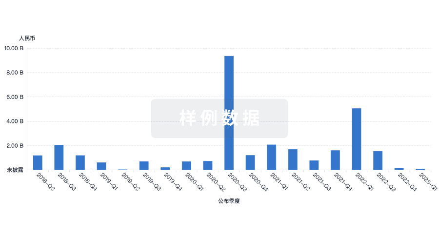
投资
深入了解从初创企业到成熟企业的最新公司投资动态。
登录
或
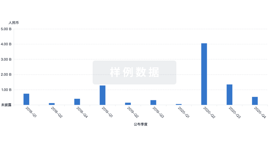
融资
发掘融资趋势以验证和推进您的投资机会。
登录
或
来和芽仔聊天吧
立即开始免费试用!
智慧芽新药情报库是智慧芽专为生命科学人士构建的基于AI的创新药情报平台,助您全方位提升您的研发与决策效率。
立即开始数据试用!
智慧芽新药库数据也通过智慧芽数据服务平台,以API或者数据包形式对外开放,助您更加充分利用智慧芽新药情报信息。
生物序列数据库
生物药研发创新
免费使用
化学结构数据库
小分子化药研发创新
免费使用