预约演示
更新于:2025-05-07
Skaggs School of Pharmacy & Pharmaceutical Sciences
更新于:2025-05-07
概览
标签
消化系统疾病
肿瘤
皮肤和肌肉骨骼疾病
小分子化药
疾病领域得分
一眼洞穿机构专注的疾病领域

技术平台
公司药物应用最多的技术
靶点
公司最常开发的靶点
关联
靶点 |
作用机制 |
在研机构 |
原研机构 |
在研适应症 |
非在研适应症 |
最高研发阶段 |
首次获批国家/地区 |
首次获批日期 |
100 项与 Skaggs School of Pharmacy & Pharmaceutical Sciences 相关的临床结果
登录后查看更多信息
登录后查看更多信息
2025-04-22Journal of Chemical Theory and Computation
Computation of Protein–Ligand Binding Free Energies with a Quantum Mechanics-Based Mining Minima Algorithm
Article
作者: Guidez, Emilie B. ; Gordon, Mark S. ; Sattasathuchana, Tosaporn ; Webb, Simon P. ; Xu, Peng ; Potter, Michael J. ; Schlinsog, Megan ; Gilson, Michael K.
2025-04-08Neurology
Acute Effects of Oral Levodopa on Seated Mean Arterial Blood Pressure in Parkinson’s Disease (P7-5.021)
作者: Litvan, Irene ; Moonla, Chochanon ; Longardner, Katherine ; Ghodsi, Hamid ; Momper, Jeremiah ; Wang, Joseph ; Mahato, Kuldeep ; Liu, Catherine
2025-04-08Analytical Chemistry
Capillary Zone Electrophoresis-Mass Spectrometry of Intact G Protein-Coupled Receptors Enables Proteoform Profiling
Article
作者: Jooß, Kevin ; Kelleher, Neil L. ; Melani, Rafael D. ; Ives, Ashley N. ; Fellers, Ryan T. ; Janetzko, John
2024-06-04
100 项与 Skaggs School of Pharmacy & Pharmaceutical Sciences 相关的药物交易
登录后查看更多信息
100 项与 Skaggs School of Pharmacy & Pharmaceutical Sciences 相关的转化医学
登录后查看更多信息
组织架构
使用我们的机构树数据加速您的研究。
登录
或

管线布局
2025年07月20日管线快照
管线布局中药物为当前组织机构及其子机构作为药物机构进行统计,早期临床1期并入临床1期,临床1/2期并入临床2期,临床2/3期并入临床3期
临床前
1
登录后查看更多信息
当前项目
登录后查看更多信息
药物交易
使用我们的药物交易数据加速您的研究。
登录
或
转化医学
使用我们的转化医学数据加速您的研究。
登录
或
营收
使用 Synapse 探索超过 36 万个组织的财务状况。
登录
或
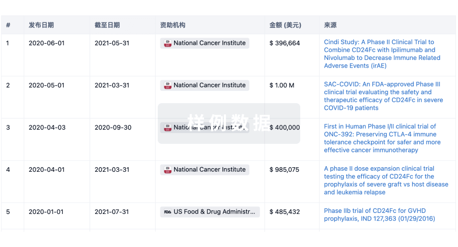
科研基金(NIH)
访问超过 200 万项资助和基金信息,以提升您的研究之旅。
登录
或
投资
深入了解从初创企业到成熟企业的最新公司投资动态。
登录
或
融资
发掘融资趋势以验证和推进您的投资机会。
登录
或
Eureka LS:
全新生物医药AI Agent 覆盖科研全链路,让突破性发现快人一步
立即开始免费试用!
智慧芽新药情报库是智慧芽专为生命科学人士构建的基于AI的创新药情报平台,助您全方位提升您的研发与决策效率。
立即开始数据试用!
智慧芽新药库数据也通过智慧芽数据服务平台,以API或者数据包形式对外开放,助您更加充分利用智慧芽新药情报信息。
生物序列数据库
生物药研发创新
免费使用
化学结构数据库
小分子化药研发创新
免费使用