预约演示
更新于:2025-11-09

University of Minho
更新于:2025-11-09
概览
标签
肿瘤
皮肤和肌肉骨骼疾病
小分子化药
疾病领域得分
一眼洞穿机构专注的疾病领域
技术平台
公司药物应用最多的技术
靶点
公司最常开发的靶点

暂无数据
关联
靶点 |
作用机制 |
在研机构 |
原研机构 |
在研适应症 |
非在研适应症 |
最高研发阶段 |
首次获批国家/地区 |
首次获批日期 |
NCT06317753
Can Exercise Rewire the Brain Addiction Circuitry? A Randomized Trial of Exercise and Binge Drinking
NCT05472194
Promoting Inclusive Education Through Executive Functions - Study Protocol of a Randomized Control Trial
NCT06823557
Gut2Brain: Investigation of the Mechanisms of the Gut-brain Axis in Binge Eating and Obesity

100 项与 University of Minho 相关的临床结果
登录后查看更多信息
登录后查看更多信息
2026-06-01Medical Gas Research
Molecular hydrogen and kidney diseases: a scoping review based on scientometry and data analytics
Article
作者: Leiva, Víctor ; Castro, Cecilia ; Viana, Johanna
Acute kidney injury and chronic kidney disease impose substantial burdens on healthcare systems worldwide. Molecular hydrogen (H2) has emerged as a potential therapy due to its selective antioxidant, anti-inflammatory, and antiapoptotic properties. The present study reviews evidence on H₂-based renal interventions, examining therapeutic mechanisms, bibliometric trends, and existing research gaps based on data analytics. This scoping review integrates quantitative bibliometric analysis with qualitative thematic synthesis. This integration, uncommon in conventional scoping reviews, reveals important gaps. Following the Preferred Reporting Items for Systematic reviews and Meta-Analyses extension for Scoping Reviews (PRISMA-ScR) guidelines, 69 publications were identified through Scopus and Web of Science. These publications mostly originated from Asia, particularly China and Japan, with clear peaks of activity in 2019 and 2024, but international collaboration remains limited. H₂ consistently demonstrated protective effects against apoptosis, fibrosis, inflammation, and oxidative stress across acute kidney injury, nephrotoxicity, transplantation, and early chronic kidney disease models. Our findings suggest that hydrogen therapy holds promise for renoprotection in both acute kidney injury and chronic kidney disease. Nonetheless, more robust clinical trials and standardized research methodologies are imperative to facilitate its broader adoption into clinical nephrology practice.
2026-02-01Biomaterials advances
Implementation of a fully biodegradable and biomimetic epicardial patch providing synergic physico-chemical, mechanical and electrical cues for myocardial infarction therapy
Article
作者: Munaron, Luca ; Giachino, Claudia ; Scarpellino, Giorgia ; Burchielli, Silvia ; Rossin, Daniela ; Lo Iacono, Marco ; Amorim, Sara ; Pires, Ricardo A ; Fiorino, Erika ; Bulgheresi, Chiara ; Sergi, Francesca ; Trouki, Cheherazade ; Cristallini, Caterina ; Rossino, Dawid ; Aubry, Matteo ; Kusmic, Claudia ; Rastaldo, Raffaella ; Vanni, Roberto ; Labardi, Massimiliano ; Barbani, Niccoletta ; Terlizzi, Domiziana ; Perveen, Sadia ; Reis, Rui L
The intrinsic limitation of myocardial tissue to self-repair after damage underscores the need for innovative approaches in addressing cardiac tissue damage post-myocardial infarction (MI). We aimed to develop an acellular, bioartificial, microstructured and electroconductive patch (PGF) made of poly(lactic-co-glycolic acid) (PLGA), Gelatin, and 9-fluorenylmethoxycarbonyl-diphenylalanine (Fmoc-FF), to foster post-MI endogenous cardiac healing capabilities. The self-assembling semi-conductive peptide Fmoc-FF was introduced to reduce the electrical impedance of the polymer components while maintaining the complete biodegradation of the patch. Unexpectedly, the electroconductive component was found to increase the patch microstructure stability, improve cardiomyoblast elongation, augment stromal cell differentiation and sustain Human induced Pluripotent Stem Cell-derived Cardiomyocytes (hiPSC-CM) beating for at least 30 days. The main outcome was demonstrated in vivo, where epicardial implantation of the PGF patch in a rat model of ischaemia-reperfusion promoted significant cardiac tissue repair: this was evidenced by preservation of the myocardial tissue, reduced fibrosis, and recruitment of endogenous c-Kit+ cells. This newly implemented patch configuration promotes efficient myocardial healing, offering a promising therapeutic approach for infarcted patients.
2026-02-01Biomaterials advances
TNF-α driven inflammation in bone tissues using an anatomical- and bio-integrated hydroxyapatite-enriched spongy-like hydrogel 3D model
Article
作者: da Silva, Lucília P ; Soares da Costa, Diana ; Correlo, Vitor M ; Bastos, Ana R ; Reis, Rui L
Inflammatory bone diseases like osteoporosis affect over 200 million people globally, yet the mechanisms by which acute inflammation progresses to chronic remain poorly understood. To address this, we developed an in vitro 3D cortical-sponge bone model of acute inflammation by introducing TNF-α. The model consists of GG-HAp spongy-like hydrogels with osteoblasts from human mesenchymal stem cells (HBM-MSCs) in an outer compartment, and human dermal microvascular endothelial cells plus supporting HBM-MSCs in an inner compartment, mimicking cortical and trabecular bone, respectively. Acute inflammation was induced by TNF-α supplementation (1, 10, or 100 ng/mL) over 7 days, and its impact on vascular assembly, osteogenesis, and inflammation was evaluated via RT-PCR, luminex and ELISA. TNF-α did not affect cell viability and minimally affected angiogenic factors release. However, 100 ng/mL of TNF-α significantly reduced type I collagen, specifically to 53 % at 3 days and to 82 % at 7 days. Pro-inflammatory cytokines (IL-1β, IL-6, IL-8, TNF-α, MCP-1, and M-CSF) gene expression and protein release were upregulated in a dose-dependent manner. For instance, 100 ng/mL of TNF-α increased IL-6 gene expression by 2.96 ± 2.38-fold at 3 days and 5.69 ± 3.09-fold at 7 days. Interestingly, 100 ng/mL of TNF-α declined IL-1β, IL-6 and TNF-α protein release from 3 to 7 days, indicating a proper resolution of the acute inflammatory response, mimicking the healing phase in bone repair. Overall, this model replicates acute inflammatory events in bone tissue, providing a quantitative platform to study TNF-α-driven bone dynamics and to evaluate targeted interventions, including anti-TNF-α therapies for osteoporosis.
2023-12-10
·今日头条
微生物疗法临床研究
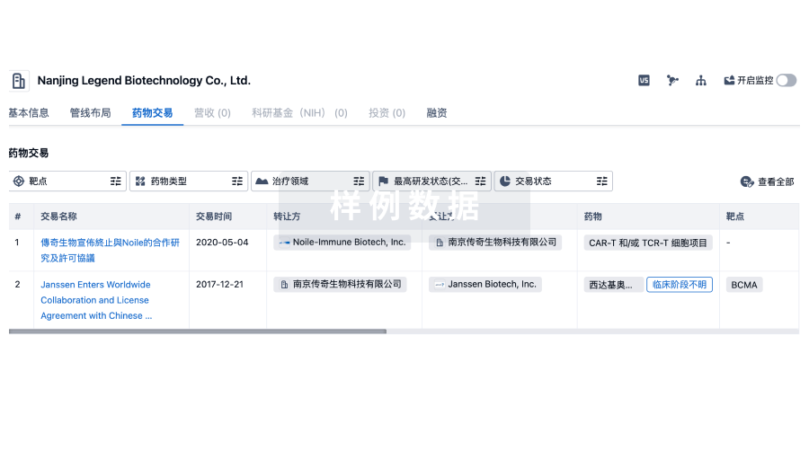
100 项与 University of Minho 相关的药物交易
登录后查看更多信息
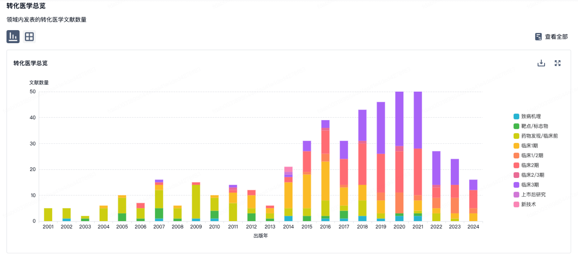
100 项与 University of Minho 相关的转化医学
登录后查看更多信息
组织架构
使用我们的机构树数据加速您的研究。
登录
或
管线布局
2025年11月10日管线快照
管线布局中药物为当前组织机构及其子机构作为药物机构进行统计,早期临床1期并入临床1期,临床1/2期并入临床2期,临床2/3期并入临床3期
临床前
1
登录后查看更多信息
当前项目
登录后查看更多信息
药物交易
使用我们的药物交易数据加速您的研究。
登录
或
转化医学
使用我们的转化医学数据加速您的研究。
登录
或
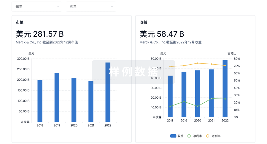
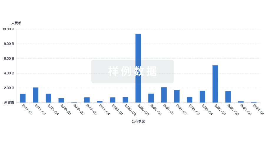
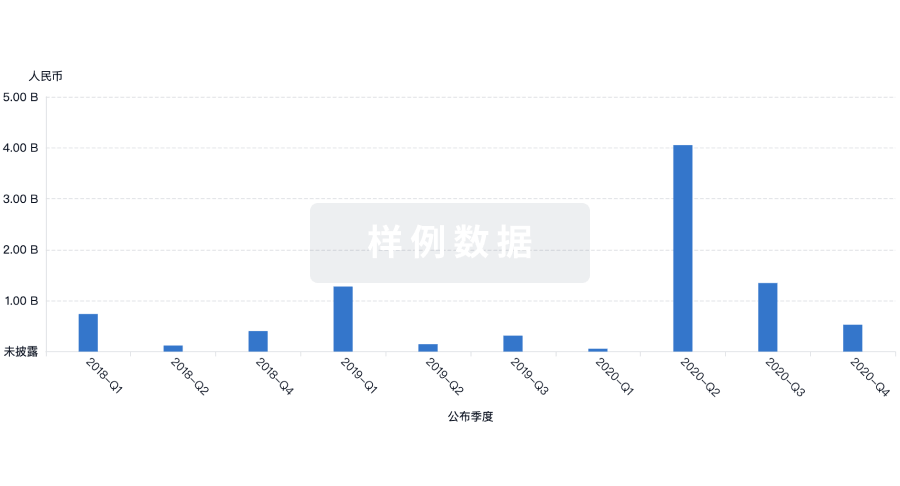
营收
使用 Synapse 探索超过 36 万个组织的财务状况。
登录
或
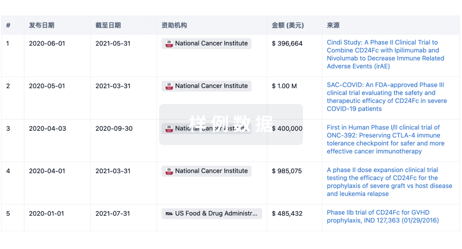
科研基金(NIH)
访问超过 200 万项资助和基金信息,以提升您的研究之旅。
登录
或
投资
深入了解从初创企业到成熟企业的最新公司投资动态。
登录
或
融资
发掘融资趋势以验证和推进您的投资机会。
登录
或
生物医药百科问答
全新生物医药AI Agent 覆盖科研全链路,让突破性发现快人一步
立即开始免费试用!
智慧芽新药情报库是智慧芽专为生命科学人士构建的基于AI的创新药情报平台,助您全方位提升您的研发与决策效率。
立即开始数据试用!
智慧芽新药库数据也通过智慧芽数据服务平台,以API或者数据包形式对外开放,助您更加充分利用智慧芽新药情报信息。
生物序列数据库
生物药研发创新
免费使用
化学结构数据库
小分子化药研发创新
免费使用