Fluorescent Hand of a Madagascar Giant Day Gecko Wins 48th Annual Nikon Small World Photo Microscopy Competition
2022-10-11
并购
The winners of the 48th annual competition showcase a spectacular blend of science and artistry under the microscope
MELVILLE, N.Y., Oct. 11, 2022 /PRNewswire/ -- Nikon Instruments Inc. today unveiled the winners of the 48th annual Nikon Small World Photomicrography Competition. This year's first place prize was awarded to Grigorii Timin, supervised by Dr. Michel Milinkovitch at the University of Geneva, for his remarkable image of an embryonic hand of a Madagascar giant day gecko. Masterfully blending imaging technology and artistic creativity, Timin utilized high-resolution microscopy and image-stitching to capture this species of Phelsuma grandis day gecko.
Continue Reading

Preview
来源: PRNewswire
Nikon Small World
Preview
来源: PRNewswire
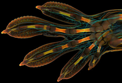
Embryonic hand of a Madagascar giant day gecko (Phelsuma grandis)
A visually stunning and painstaking technique, Timin used image-stitching to merge hundreds of images together to create the final image of his gecko. Preparing the sample was an added challenge. Timin performed whole-mount fluorescent staining and tissue clearing to capture the entire embryonic hand with a confocal microscope.
"This embryonic hand is about 3 mm (0.12 in) in length, which is a huge sample for high-resolution microscopy," said Timin. "The scan consists of 300 tiles, each containing about 250 optical sections, resulting in more than two days of acquisition and approximately 200 GB of data."
The final result gives a glimpse into the hidden beauty and complexity of the gecko, highlighting the nerves in a cyan color and the bones, tendons, ligaments, skin and blood cells in a range of warmer colors. "This particular image is beautiful and informative, as an overview and also when you magnify it in a certain region, shedding light on how the structures are organized on a cellular level," said Timin.
He went on to say, "The Nikon Small World Competition is a great opportunity to share how impressive nature is on a microscopic level, not only within a scientific community but also with the general public."
"Each year, Nikon Small World receives an array of microscopic images that exhibit exemplary scientific technique and artistry. This year was no exception," said Eric Flem, Communications and CRM Manager, Nikon Instruments. "At the intersection of art and science, this year's competition highlights stunning imagery from scientists, artists, and photomicrographers of all experience levels and backgrounds from across the globe."
Second place was awarded to Dr. Caleb Dawson for his image of breast tissue showing contractile myoepithelial cells wrapped around milk-producing alveoli. Taking a week to process, the myoepithelial cells were stained with multiple rounds of fluorescent dyes and captured with a confocal microscope.
Third place was captured by Satu Paavonsalo and Dr. Sinem Karaman for their image of blood vessel networks in the intestine of an adult mouse.
In addition to the top three winners, Nikon Small World recognized 89 photos out of thousands of entries from scientists and artists across the globe.
The 2022 judging panel included:
Dr. Gustavo Menezes, Associate Professor and Head of the Center for Gastrointestinal Biology at the Federal University of Minas Gerais
Dr. Nikolay Nikolov, Senior Video Journalist at The New York Times
Annaliese Nurnberg, Photo Editor at The Washington Post
Dr. Clare Waterman, Cell Biologist and Member of the National Academy of Sciences
For additional information, please visit www.nikonsmallworld.com, or follow the conversation on Facebook, Twitter @NikonSmallWorld and Instagram @NikonInstruments.
NIKON SMALL WORLD WINNERS
Grigorii Timin & Dr. Michel Milinkovitch
Department of Genetics and Evolution
Geneva, Switzerland
Embryonic hand of a Madagascar giant day gecko (Phelsuma grandis)
Confocal
63X (Objective Lens Magnification)
Dr. Caleb Dawson
WEHI, The Walter and Eliza Hall Institute of Medical Research
Department of Immunology
Melbourne, Victoria, Australia
Breast tissue showing contractile myoepithelial cells wrapped around milk-producing alveoli
Confocal
40X (Objective Lens Magnification)
Satu Paavonsalo & Dr. Sinem Karaman
Individualized Drug Therapy Research Program, Faculty of Medicine
Helsinki, Finland
Blood vessel networks in the intestine of an adult mouse
Confocal
10X (Objective Lens Magnification)
4th Place
Dr. Andrew Posselt
University of California, San Francisco (UCSF)
Department of Surgery
Mill Valley, California, USA
Long-bodied cellar/daddy long-legs spider (Pholcus phalangioides)
Image Stacking
3X (Objective Lens Magnification)
5th Place
Alison Pollack
San Anselmo, California, USA
Slime mold (Lamproderma)
Image Stacking, Reflected Light
10X (Objective Lens Magnification)
Ole Bielfeldt
Macrofying
Cologne, North Rhine-Westphalia, Germany
Unburned particles of carbon released when the hydrocarbon chain of candle wax breaks down
Brightfield, Image Stacking
2.5X (Objective Lens Magnification)
Dr. Jianqun Gao & Prof. Glenda Halliday
Central Clinical School / Professor Glenda Halliday's Lab
Sydney, New South Wales, Australia
Human neurons derived from neural stem cells (NSCs)
Confocal, Fluorescence
20X (Objective Lens Magnification)
8th Place
Dr. Nathanaël Prunet
University of North Carolina at Chapel Hill
Department of Biology
Chapel Hill, North Carolina, USA
Growing tip of a red algae
Confocal
10X (Objective Lens Magnification)
9th Place
Dr. Marek Sutkowski
Institute of Microelectronics and Optoelectronics
Warsaw, Poland
Liquid crystal mixture (smectic Felix 015)
Image Stacking, Polarized Light
40X (Objective Lens Magnification)
10th Place
Murat Öztürk
Ankara, Turkey
A fly under the chin of a tiger beetle
Image Stacking
3.7X (Objective Lens Magnification)
11th Place
Ye Fei Zhang
Jiang Yin, Jiangsu, China
Moth eggs
Image Stacking
10X (Objective Lens Magnification)
12th Place
Brett M. Lewis
Department of Earth and Atmospheric Science
Brisbane, Queensland, Australia
Autofluorescence of a single coral polyp (approx. 1 mm)
Fluorescence, Image Stacking
20X (Objective Lens Magnification)
13th Place
Randy Fullbright
Fullbright Studio
Vernal, Utah, USA
Agatized dinosaur bone
Image Stacking
60X (Objective Lens Magnification)
14th Place
Nadia Efimova
Philadelphia, Pennsylvania, USA
Differentiated cultured mouse myoblasts with lysosomes (cyan/green), nuclei (yellow), F-actin (magenta)
Confocal
40X (Objective Lens Magnification)
15th Place
Dr. Ziad El-Zaatari
Houston Methodist Hospital
Department of Pathology and Genomic Medicine
Houston, Texas, USA
Cross sections of normal human colon epithelial crypts
Brightfield
20X (Objective Lens Magnification)
16th Place
Dr. Olivier Leroux
Department of Biology & Department of Plants and Crops
Ghent, Oost-Vlaanderen, Belgium
Longitudinal section through a white asparagus shoot tip
10X (Objective Lens Magnification)
17th Place
Dr. Daniel Wehner & Julia Kolb
Max Planck Institute for the Science of Light
Department of Biological Optomechanics
Erlangen, Bavaria, Germany
Tail fin of a zebrafish larva with peripheral nerves (green) and extracellular matrix (violet)
Confocal
10X (Objective Lens Magnification)
Dr. Julien Resseguier
Department of Biosciences / Immunology
Oslo, Viken, Norway
Network of macrophages (white blood cells) of an adult zebrafish intestine
Confocal, Deconvolution, Fluorescence
60X (Objective Lens Magnification)
19th Place
Dr. Tagide deCarvalho
University of Maryland, Baltimore County (UMBC)
Keith R. Porter Imaging Facility
Baltimore, Maryland, USA
Bacterial biofilm on a human tongue cell
Confocal
63X (Objective Lens Magnification)
20th Place
Hui Lin & Dr. Kim McBride
Nationwide Children's Hospital
Center for Cardiovascular Research
Columbus, Ohio, USA
Human cardiomyocytes (heart cells) derived from induced pluripotent stem cells
60X (Objective Lens Magnification)
HONORABLE MENTIONS
Dr. Dylan T. Burnette
Department of Cell and Developmental Biology
Nashville, Tennessee, USA
A crawling cell
Structured Illumination Microscopy (SIM)
60X (Objective Lens Magnification)
Dr. Amy C. Engevik
Department of Regenerative Medicine & Cell Biology
Charleston, South Carolina, USA
Intestinal villi (brush border in magenta)
20X (Objective Lens Magnification)
Dr. Laurent Formery
Department of Molecular and Cell Biology
Pacific Grove, California, USA
Two-month old juvenile sea star (Patiria miniata)
Confocal, Fluorescence, Image Stacking
20X (Objective Lens Magnification)
Karl Gaff
Dublin, Ireland
Midge larva collected from a fresh water pond
Polarized Light
10X (Objective Lens Magnification)
Gerd Günther
Düsseldorf, Germany
Young stem of garden bamboo (Fargesia sp.)
10X (Objective Lens Magnification)
Dr. Zhiguo He
University Jean Monnet
School of Medicine
Saint-Priest-en-Jarez, Rhône-Alpes, France
The actomyosin network at the apical pole of human corneal endothelial cells (revealed by immunofluorescence)
60X (Objective Lens Magnification)
Department of Biology
Philadelphia, Pennsylvania, USA
Migrating human fibroblast stained for the golgi (orange), the actin cytoskeleton (magenta), and the nucleus (cyan)
Confocal, Fluorescence
60X (Objective Lens Magnification)
Reuben Philip
Mount Sinai Hospital
Toronto, Ontario, Canada
A cell with extra centrosomes beginning to divide
Confocal
60X (Objective Lens Magnification)
Alison Pollack
San Anselmo, California, USA
Slime mold (Didymium clavus)
Image Stacking, Reflected Light
10X (Objective Lens Magnification)
Jan Rosenboom
Rostock, Mecklenburg Vorpommern, Germany
Diatom (Actinoptychus sp.)
Differential Interference Contrast (DIC), Image Stacking
100X (Objective Lens Magnification)
Dr. Igor Siwanowicz
Howard Hughes Medical Institute (HHMI)
Janelia Research Campus
Ashburn, Virginia, USA
Radula (rasping tongue) of a marine snail (Turbinidae family)
Confocal
10X (Objective Lens Magnification)
Sebastian Sparenga
Chicago, Illinois, USA
Recrystallized Vitamin C
Polarized Light
10X (Objective Lens Magnification)
Dr. Andrea Tedeschi
The Ohio State University / Wexner Medical Center
Department of Neuroscience
Columbus, Ohio, USA
Murine sensory-motor cortex following mild traumatic brain injury in a transgenic mouse (expressing Thy1-GFP)
Confocal
10X (Objective Lens Magnification)
Wim van Egmond
Micropolitan Museum
Berkel en Rodenrijs, Zuid-Holland, Netherlands
Larva of an anemone, found in marine plankton
Darkfield
6.3X (Objective Lens Magnification)
Ye Fei Zhang
Jiang Yin, Jiangsu, China
Butterfly egg
Image Stacking
10X (Objective Lens Magnification)
About Nikon Small World Photomicrography Competition
The Nikon Small World Photomicrography Competition is open to anyone with an interest in photography or video. Participants may upload digital images and videos directly at www.nikonsmallworld.com. For additional information, contact Nikon Small World, Nikon Instruments Inc., 1300 Walt Whitman Road, Melville, NY 11747, USA, or phone (631) 547-8569. Entry forms for Nikon's 2023 Small World and Small World in Motion Competitions are available at https://enter.nikonsmallworld.com/
About Nikon Instruments Inc.
Nikon Instruments Inc. is the US microscopy arm of Nikon Healthcare, a world leader in the development and manufacture of optical and digital imaging technology for biomedical applications. For more information, visit https://www.microscope.healthcare.nikon.com/ or contact us at 1-800-52-NIKON.
SOURCE Nikon Instruments Inc.
更多内容,请访问原始网站
文中所述内容并不反映新药情报库及其所属公司任何意见及观点,如有版权侵扰或错误之处,请及时联系我们,我们会在24小时内配合处理。
适应症
靶点
-药物
-热门报告


立即开始免费试用!
智慧芽新药情报库是智慧芽专为生命科学人士构建的基于AI的创新药情报平台,助您全方位提升您的研发与决策效率。
立即开始数据试用!
智慧芽新药库数据也通过智慧芽数据服务平台,以API或者数据包形式对外开放,助您更加充分利用智慧芽新药情报信息。