预约演示
更新于:2025-08-29
Stuart Pharmaceuticals
子公司|United States
子公司|United States
更新于:2025-08-29
概览
关联
100 项与 Stuart Pharmaceuticals 相关的临床结果
登录后查看更多信息
0 项与 Stuart Pharmaceuticals 相关的专利(医药)
登录后查看更多信息
7
项与 Stuart Pharmaceuticals 相关的文献(医药)1988-06-01·American journal of veterinary research3区 · 农林科学
Comparison of clinical judgment, Doppler ultrasound, and fluorescein fluorescence as methods for predicting intestinal viability in the pony
3区 · 农林科学
Article
作者: Freeman, David E. ; Fetrow, John P. ; Cimprich, Ronnie ; Richardson, Dean W. ; Orsini, James A. ; Tulleners, Eric P. ; Gentile, Dennis G.
SUMMARY:
Strangulation obstruction was induced in anesthetized ponies for periods of 2 and 3 hours by clamping 45-cm segments of jejunum and associated veins (venous strangulation obstruction) and arteries and veins (arterial and venous strangulation obstruction). Four segments were studied in each of 7 ponies allowed to survive 12 hours, 2 segments in a pony that was allowed to survive 1 hour, and 1 segment in each of 10 ponies allowed to survive 42 days after the strangulation periods ended. Fifteen minutes after the periods of strangulation obstruction ended, the viability of test segments was assessed by clinical judgment (40 segments), fluorescein fluorescence (40 segments), and Doppler ultrasound (32 segments). Because the test segments were normal at necropsy in long-term survivors, all segments were designated as viable.The overall accuracy of the methods used to predict viability was 88% for Doppler ultrasound and 53% each for clinical judgment and fluorescein fluorescence (P < 0.005). Failures in the last 2 techniques could be attributed to their tendency to score venous strangulation obstruction segments as nonviable (90% for each). Doppler ultrasound was 94% accurate in these segments.
1988-05-01·American journal of veterinary research3区 · 农林科学
Influence of nonbiologic implants on laminectomy membrane formation in dogs
3区 · 农林科学
Article
作者: Crissman, J. ; Babish, J. ; Trotter, E. J. ; Robson, D.
SUMMARY:
The effects of various surgical implants, spinal cord hypothermia, and glucocorticoid administration on formation of the laminectomy membrane were evaluated in 32 preconditioned chondrodystrophoid dogs. Modified dorsal laminectomies and full-length durotomies, from T12 to L1, were performed on all dogs. Dogs were allotted to 2 groups. Group-1 dogs (n = 20) were further allocated to 4 subgroups (a, b, c, and d) consisting of 5 dogs each. Group-1a dogs received no implant, group-1b dogs had absorbable gelatin sponges implanted, group-1c dogs had absorbable gelatin films implanted, and group-1d dogs had absorbable gelatin sponges and absorbable gelatin films implanted. Daily neurologic examinations permitted correlation of neurologic dysfunction with secondary spinal cord compression in those dogs in which it developed. The influence of these implants on laminectomy membrane formation and dural healing was assessed by gross and microscopic evaluation of transverse sections of the vertebrae and spinal cord after euthanasia of one member of each subgroup at 1, 2, 4, 8, and 16 weeks after surgery.Group-2 dogs (n = 12) were further allotted to 3 subgroups (a, b, and c) consisting of 4 dogs each. One dog in each group-2 subgroup underwent the same surgical procedures described for the group-1 subgroups (ie, 4 procedures/group-2 subgroup). The additional effects of 3 conventional supportive techniques (selective regional spinal cord hypothermia, glucocorticoid administration, or spinal cord hypothermia and glucocorticoid administration) on laminectomy membrane formation and on immediate postoperative recovery were examined in groups 2a, 2b, and 2c, respectively. Neurologic examinations were performed daily until this time. All dogs in group 2 were euthanatized 1 week after surgery for gross and microscopic examination of transverse sections of the vertebrae and spinal cord.Qualitative histopathologic effects of the different implants and supportive techniques on formation of the laminectomy membrane were determined. Statistical analysis of the degrees of secondary spinal cord compression was performed in group-1 dogs by measuring and comparing ratios of the vertical to the horizontal diameters or the transverse spinal cord sections from locations within (T12 to L1 and out of (T11, T11-12, L1-2, and L2) the region of surgical intervention. The vertical/horizontal diameter ratios measured from transverse sections from T11 to L2 in size-matched, untreated control dogs formed the standards for a mean roundness index of the spinal cord in the various anatomic locations of the vertebral column.A significant difference was not detected between measurements from dogs in which there was no implantation and those from dogs in which absorbable gelatin sponge was implanted, or between measurements from dogs receiving absorbable gelatin film and those from dogs receiving both absorbable gelatin sponge and absorbable gelatin film. There were, however, significant (P < 0.05) differences between measurements from dogs that did not receive an implant and those from dogs in which absorbable gelatin film was implanted, between dogs that did not receive an implant and those in which absorbable gelatin sponge/absorbable gelatin film was implanted, between dogs in which absorbable gelatin sponge was implanted and those in which absorbable gelatin film was implanted, and between dogs in which absorbable gelatin sponge was implanted and dogs in which absorbable gelatin sponge/absorbable gelatin film was implanted. No implant or absorbable gelatin sponge implantation resulted in less compression of the spinal cord than did the other implants.The ancillary supportive techniques resulted in readily identifiable microscopic changes in the healing patterns of the laminectomy defects, but did not significantly alter the spinal cord compression or the transient postoperative neurologic deficits.
1988-05-01·American journal of surgery3区 · 医学
Analysis of prothrombin time prolongation in north American cefotetan clinical trials: Questions and answers
3区 · 医学
Article
作者: Goldstein, N H
100 项与 Stuart Pharmaceuticals 相关的药物交易
登录后查看更多信息
100 项与 Stuart Pharmaceuticals 相关的转化医学
登录后查看更多信息
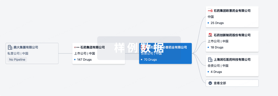
组织架构
使用我们的机构树数据加速您的研究。
登录
或
管线布局
2025年11月05日管线快照
管线布局中药物为当前组织机构及其子机构作为药物机构进行统计,早期临床1期并入临床1期,临床1/2期并入临床2期,临床2/3期并入临床3期
其他
1
登录后查看更多信息
当前项目
| 药物(靶点) | 适应症 | 全球最高研发状态 |
|---|---|---|
盐酸布克力嗪 ( H1 receptor ) | 恶心 更多 | 撤市 |
登录后查看更多信息
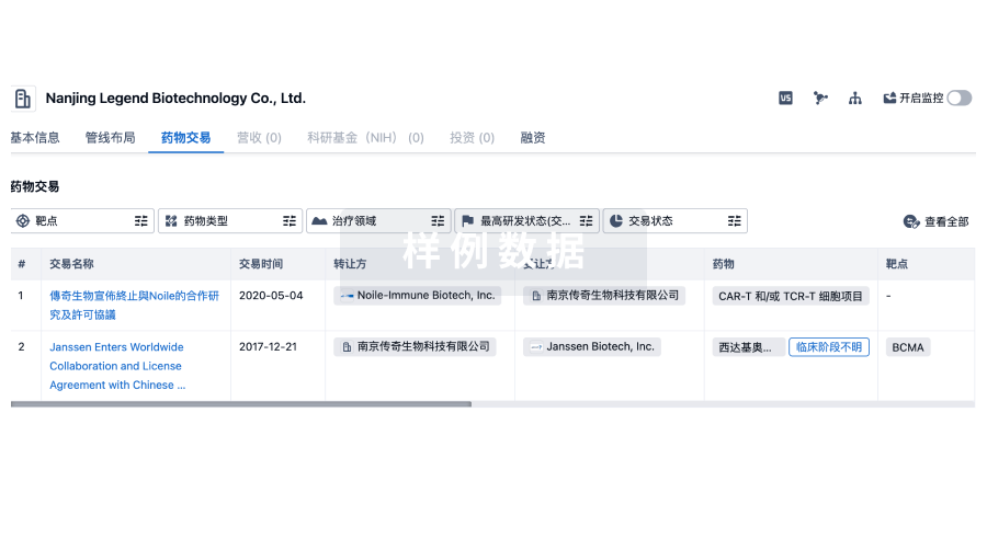
药物交易
使用我们的药物交易数据加速您的研究。
登录
或
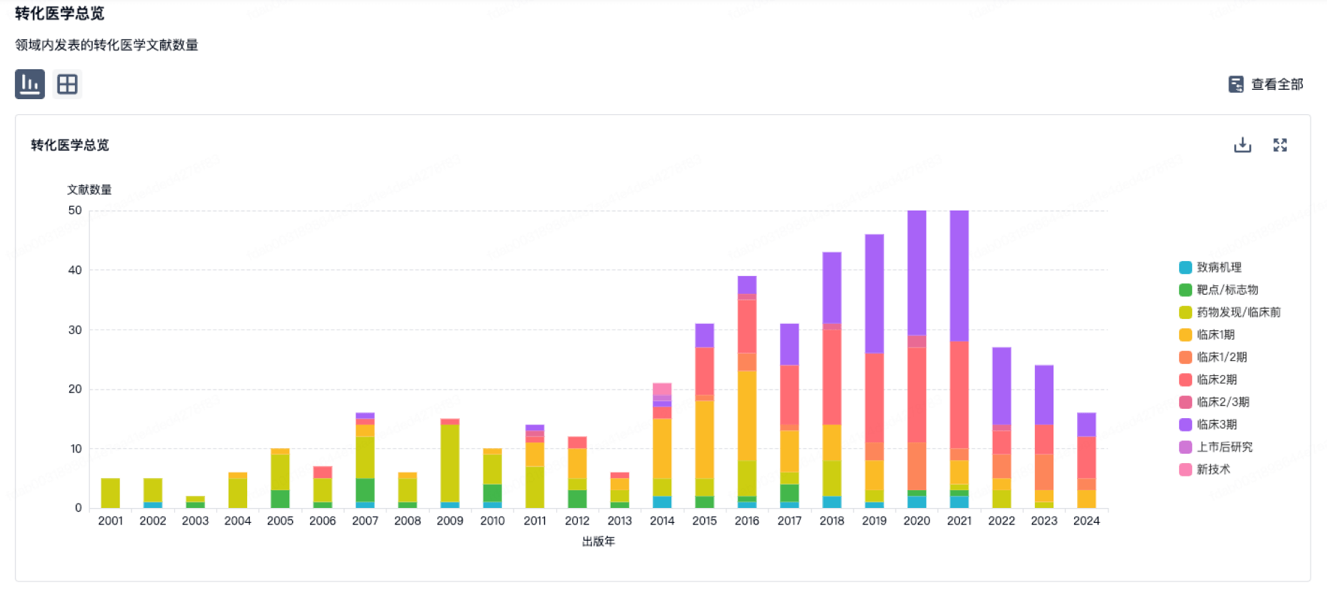
转化医学
使用我们的转化医学数据加速您的研究。
登录
或
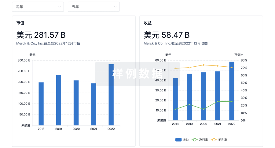
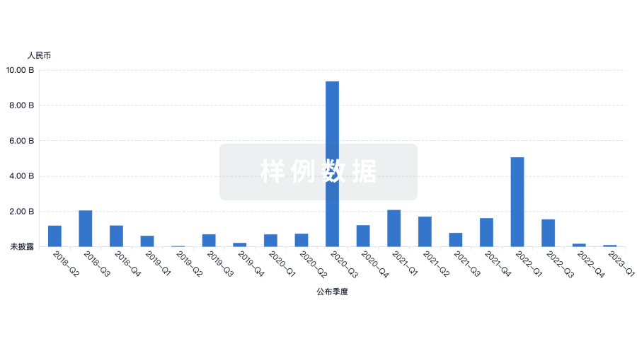
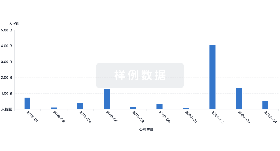
营收
使用 Synapse 探索超过 36 万个组织的财务状况。
登录
或
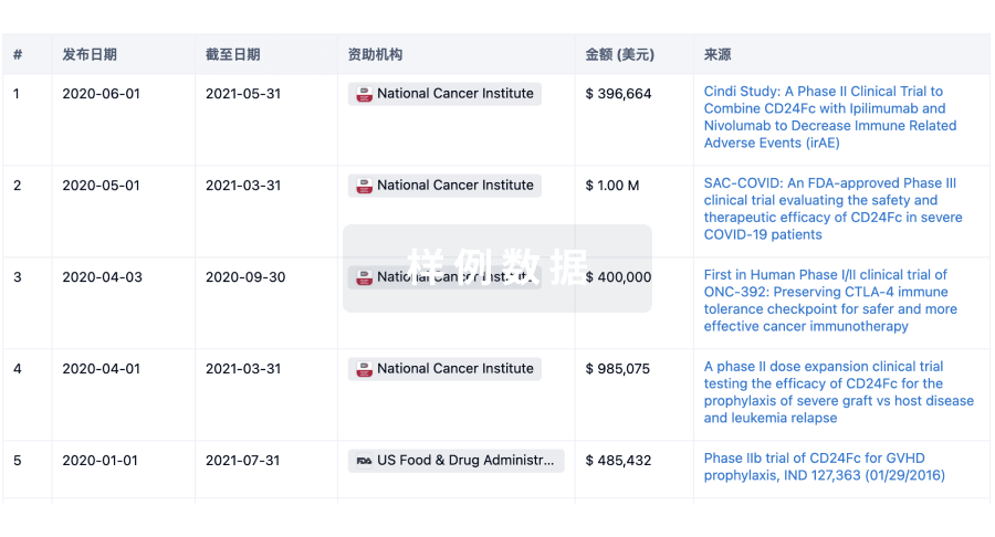
科研基金(NIH)
访问超过 200 万项资助和基金信息,以提升您的研究之旅。
登录
或
投资
深入了解从初创企业到成熟企业的最新公司投资动态。
登录
或
融资
发掘融资趋势以验证和推进您的投资机会。
登录
或
生物医药百科问答
全新生物医药AI Agent 覆盖科研全链路,让突破性发现快人一步
立即开始免费试用!
智慧芽新药情报库是智慧芽专为生命科学人士构建的基于AI的创新药情报平台,助您全方位提升您的研发与决策效率。
立即开始数据试用!
智慧芽新药库数据也通过智慧芽数据服务平台,以API或者数据包形式对外开放,助您更加充分利用智慧芽新药情报信息。
生物序列数据库
生物药研发创新
免费使用
化学结构数据库
小分子化药研发创新
免费使用