预约演示
更新于:2025-04-06
Datun Coal & Electricity (Group) Co., Ltd.
更新于:2025-04-06
概览
关联
100 项与 大屯煤电(集团)有限责任公司 相关的临床结果
登录后查看更多信息
0 项与 大屯煤电(集团)有限责任公司 相关的专利(医药)
登录后查看更多信息
2
项与 大屯煤电(集团)有限责任公司 相关的文献(医药)2014-12-01·Xi bao yu fen zi mian yi xue za zhi = Chinese journal of cellular and molecular immunology
[Expression and significance of hypoxia-inducible factor-1α in lung tissues of obesity-asthma rat].
Article
作者: Shi, Ruirui ; Chen, Lin ; Zhu, Shuyang ; Zhu, Jiechen ; Pei, Houshuang
OBJECTIVE:
To evaluate the expression level of hypoxia-inducible factor-1α (HIF-1α) in the rat model of diet-induced obesity and asthma.
METHODS:
Forty male specific pathogen-free SD rats were randomly divided into four groups: normal body mass control group (group A), asthmatic rats with normal body mass (group B), obese control group (group C) and obese asthmatic rats (group D). The rats in both group A and B were fed basic diet, while those in group C and D were fed high-fat diet in order to establish diet-induced obese rat model. Rats in group B and D were sensitized and challenged with chicken ovalbumin (OVA) to establish the asthmatic model. The white cell count in bronchoalveolar lavage fluid (BALF) was performed. The total area of the airway wall (Wat) was measured by ImagePro Plus software and was standardized by the basement membrane perimeter (Pbm). The expression of HIF-1α in lung tissue was detected by immunohistochemistry. The concentration of HIF-1α in serum was determined by ELISA. The relationships of the total white cells in BALF and airway wall thickness (WAt/Pbm) with the expression of HIF-1α were analyzed by Pearson correlation analysis.
RESULTS:
The total number of white cells in BALF in group D was (98.0±5.5)×10(4)/mL, which was significantly higher than those in group A (24.7±3.3)×10(4)/mL, group C (26.1±3.8)×10(4)/mL and group B (87.8±7.1)×10(4)/mL. The thickness of airway wall (WAt/Pbm) in group D was (9.91±0.56)m(2)/m, which was significantly higher than those in group A (6.11±0.99)m(2)/m and group C(5.99±0.83)m(2)/m, but when compared with that in group B (8.60±0.53)m(2)/m, there was no significant difference. The percentage of HIF-1α positive cells in group D was (19.44±0.96)%, which was significantly higher than those in group A (2.19±0.91)%, group C(2.56±0.89)% and group B (18.25±1.29)% (all P<0.05). The expression of HIF-1α in blood serum and BALF in group D were respectively(29.107±1.576) ng/mL and (0.511±0.011) ng/mL, which were significantly higher than those in group A [(19.380±1.506) ng/mL and (0.280±0.008) ng/mL, respectively], group C [(20.782±2.034) ng/mL and (0.281±0.010) ng/mL, respectively] and group B [(23.961±1.565) ng/mL and (0.397±0.011) ng/mL, respectively]. The expression of HIF-1α was positively correlated with the total number of white cells in BALF and the airway wall thickness.
CONCLUSION:
The expression of HIF-1α in serum and lung tissue from obese asthmatic rats significantly increases, which is positively correlated with the total number of white cells in BALF and the airway wall thickness.
Zhiye Yu Jiankang
Health status of liver among underground tunnelling workers in Datun coal mine
作者: Meng, Jie ; Fan, Jing-jing ; Ni, Chun-hui ; Wang, Shu-ping ; Luo, Chen
The health status of liver among 895 underground tunnelling workers of Datun coal mine (Xuzhuang mine and Yaoqiao mine) was investigated and analyzed, problems were tried to find out, and scientific evidence for further improving the health protection measures and health care strategy was provided.The self-designed "dust-exposed workers questionnaire" was used to investigate 895 tunnelling workers of Datun coal mine.Ultrasonic examination was done by Datun coal mine hospital physician, the levels of hepatic biochem. indexes were detected by the hospital clin. laboratoryThe Epidata 31 software was used to form the database and State100 software was applied for data anal.The detection rate of fatty liver was 29.05%, and the abnormal rate of alanine aminotransferase (ALT) was 8.49%.The detection rates of ALT, total bilirubin (TBIL) and direct bilirubin (DBIL) in Yaoqiao mine were significantly higher than those of the Xuzhuang mine, as well as fatty liver and liver cyst (P<0.01).Regular drinking was one of the main independent risk factors of fatty liver.The fatty liver is one of the main liver diseases in Datun coal mines.The high detection rate of fatty liver and other liver diseases has a certain relationship with workers living habits.
100 项与 大屯煤电(集团)有限责任公司 相关的药物交易
登录后查看更多信息
100 项与 大屯煤电(集团)有限责任公司 相关的转化医学
登录后查看更多信息
组织架构
使用我们的机构树数据加速您的研究。
登录
或
管线布局
2025年05月06日管线快照
无数据报导
登录后保持更新
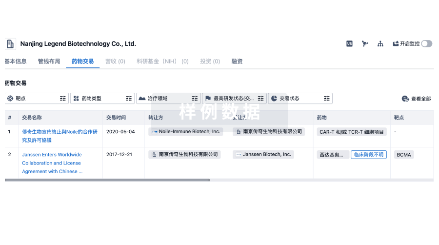
药物交易
使用我们的药物交易数据加速您的研究。
登录
或
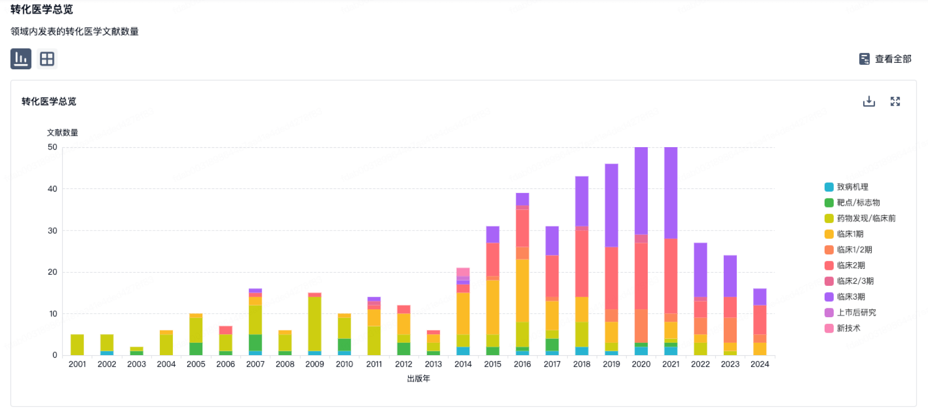
转化医学
使用我们的转化医学数据加速您的研究。
登录
或
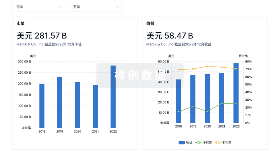
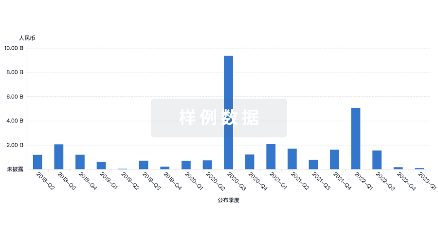
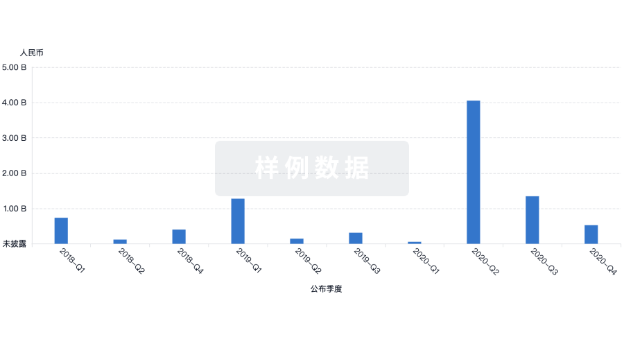
营收
使用 Synapse 探索超过 36 万个组织的财务状况。
登录
或
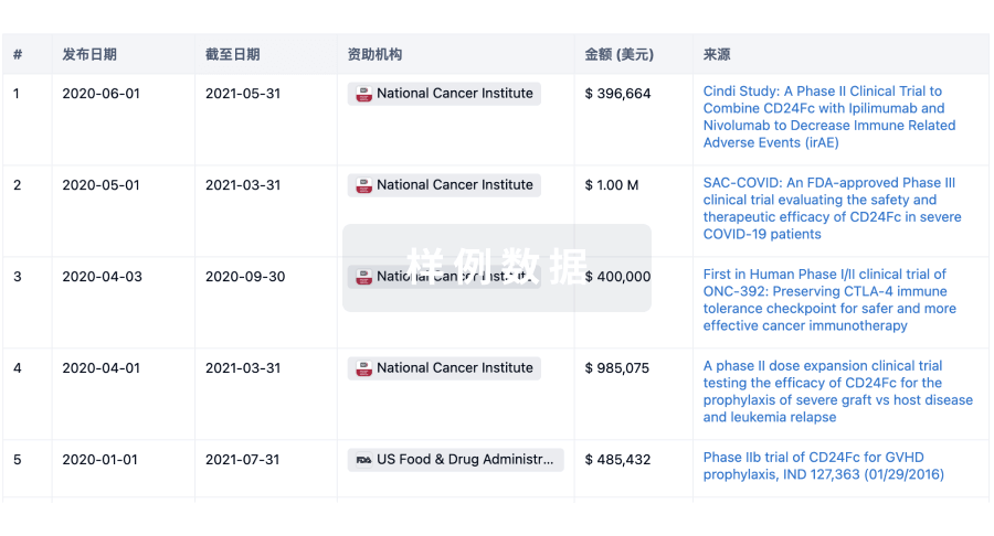
科研基金(NIH)
访问超过 200 万项资助和基金信息,以提升您的研究之旅。
登录
或
投资
深入了解从初创企业到成熟企业的最新公司投资动态。
登录
或
融资
发掘融资趋势以验证和推进您的投资机会。
登录
或
Eureka LS:
全新生物医药AI Agent 覆盖科研全链路,让突破性发现快人一步
立即开始免费试用!
智慧芽新药情报库是智慧芽专为生命科学人士构建的基于AI的创新药情报平台,助您全方位提升您的研发与决策效率。
立即开始数据试用!
智慧芽新药库数据也通过智慧芽数据服务平台,以API或者数据包形式对外开放,助您更加充分利用智慧芽新药情报信息。
生物序列数据库
生物药研发创新
免费使用
化学结构数据库
小分子化药研发创新
免费使用