预约演示
更新于:2025-06-13
GE Medical Systems Israel Ltd.
子公司|Israel
子公司|Israel
更新于:2025-06-13
概览
关联
100 项与 GE Medical Systems Israel Ltd. 相关的临床结果
登录后查看更多信息
0 项与 GE Medical Systems Israel Ltd. 相关的专利(医药)
登录后查看更多信息
3
项与 GE Medical Systems Israel Ltd. 相关的文献(医药)2017-12-01·Journal of therapeutic ultrasound
A focused ultrasound treatment system for moving targets (part I): generic system design and in-silico first-stage evaluation
Article
作者: Günther, Matthias ; Mihcin, Senay ; Barrios Romero, Diego ; Preusser, Tobias ; Jenne, Jürgen ; Bezzi, Mario ; Zidowitz, Stephan ; Sat, Giora ; Haase, Sabrina ; Levy, Yoav ; Tanner, Christine ; Schwenke, Michael ; von Dresky, Caroline ; Rothlübbers, Sven ; Strehlow, Jan ; Melzer, Andreas ; Georgii, Joachim ; Demedts, Daniel
BACKGROUND:
Focused ultrasound (FUS) is entering clinical routine as a treatment option. Currently, no clinically available FUS treatment system features automated respiratory motion compensation. The required quality standards make developing such a system challenging.
METHODS:
A novel FUS treatment system with motion compensation is described, developed with the goal of clinical use. The system comprises a clinically available MR device and FUS transducer system. The controller is very generic and could use any suitable MR or FUS device. MR image sequences (echo planar imaging) are acquired for both motion observation and thermometry. Based on anatomical feature tracking, motion predictions are estimated to compensate for processing delays. FUS control parameters are computed repeatedly and sent to the hardware to steer the focus to the (estimated) target position. All involved calculations produce individually known errors, yet their impact on therapy outcome is unclear. This is solved by defining an intuitive quality measure that compares the achieved temperature to the static scenario, resulting in an overall efficiency with respect to temperature rise. To allow for extensive testing of the system over wide ranges of parameters and algorithmic choices, we replace the actual MR and FUS devices by a virtual system. It emulates the hardware and, using numerical simulations of FUS during motion, predicts the local temperature rise in the tissue resulting from the controls it receives.
RESULTS:
With a clinically available monitoring image rate of 6.67 Hz and 20 FUS control updates per second, normal respiratory motion is estimated to be compensable with an estimated efficiency of 80%. This reduces to about 70% for motion scaled by 1.5. Extensive testing (6347 simulated sonications) over wide ranges of parameters shows that the main source of error is the temporal motion prediction. A history-based motion prediction method performs better than a simple linear extrapolator.
CONCLUSIONS:
The estimated efficiency of the new treatment system is already suited for clinical applications. The simulation-based in-silico testing as a first-stage validation reduces the efforts of real-world testing. Due to the extensible modular design, the described approach might lead to faster translations from research to clinical practice.
2016-06-01·International journal of computer assisted radiology and surgery3区 · 工程技术
In vivo validation of spatio-temporal liver motion prediction from motion tracked on MR thermometry images
3区 · 工程技术
Article
作者: Melzer, A ; Houston, G ; Preusser, T ; Tanner, C ; Zur, Y ; French, K ; Samei, G ; Strehlow, J ; Kozerke, S ; Sat, G ; McLeod, H ; Székely, G
PURPOSE:
Magnetic resonance-guided focused ultrasound (MRgFUS) of the liver during free-breathing requires spatio-temporal prediction of the liver motion from partial motion observations. The study purpose is to evaluate the prediction accuracy for a realistic MRgFUS therapy scenario, namely for human in vivo data, tracking based on MR images routinely acquired during MRgFUS and in vivo deformations caused by the FUS probe.
METHODS:
In vivo validation of the motion model was based on a 3D breath-hold image and an interleaved acquisition of two MR slices. Prediction accuracy was determined with respect to manually annotated landmarks. A statistical population liver motion model was used for predicting the liver motion for not tracked regions. This model was individualized by mapping it to end-exhale 3D breath-hold images. Spatial correspondence between tracking and model positions was established by affine 3D-to-2D image registration. For spatio-temporal prediction, MR tracking results were temporally extrapolated.
RESULTS:
Performance was evaluated for 10 volunteers, of which 5 had a dummy FUS probe put on their abdomen. MR tracking had a mean (95 %) accuracy of 1.1 (2.4) mm. The motion of the liver on the evaluation MR slice was spatio-temporally predicted with an accuracy of 1.9 (4.4) mm for a latency of 216 ms. A simple translation model performed similarly (2.1 (4.8) mm) as the two MR slices were relatively close (mean 38 mm). Temporal prediction was important (10 % error reduction), while registration effects could only partially be assessed and showed no benefits. On average, motion magnitude, motion amplitude and breathing frequency increased by 24, 16 and 8 %, respectively, for the cases with FUS probe placement. This motion increase could be reduced by the spatio-temporal prediction.
CONCLUSION:
The study shows that tracking liver vessels on MR images, which are also used for MR thermometry, is a viable approach.
2000-03-01·Magnetic resonance in medicine2区 · 医学
Design of improved spectral-spatial pulses for routine clinical use
2区 · 医学
Article
作者: Zur, Y
Spectral-spatial pulses (spsp pulses) selectively excite spins at spatial location z and spectral frequency (due to chemical shift and/or field inhomogeneity) v. In this work we discuss the design of improved spsp pulses for fat signal suppression. Optimal pulses are designed as optimal constant ripple FIR filters using the inverse SLR transform. Spsp pulses with thin slices are obtained by modifying the phases between subpulses, thereby eliminating unwanted magnetization lobes. Robust spsp pulses at off-center slices are obtained with a prescan calibration. These pulses are used either for selective fat saturation or for selective water excitation. It is shown that spsp pulses suppress fat signal better than conventional fat saturation pulses. Using the techniques presented in this article, we replaced all the fat saturation pulses on our systems with spsp pulses and obtained a significant improvement in image quality.
149
项与 GE Medical Systems Israel Ltd. 相关的新闻(医药)2025-06-12
关注并星标CPHI制药在线 近日,强生医疗科技内部公告称,中国区副总裁、首席数字官戴鹰将于6月30日正式离职,戴鹰表示离开强生出于个人选择,目的为了"寻求外部发展机会",强生中国研发团队负责人Joanna Wan将暂时肩负起数字化团队的领导职责。戴鹰1998年加入GE医疗,24年间从中国区工程部经理一路升至全球研发中心总经理,主导了GE医疗中国的国产化战略与数字化转型。2022年加入强生后,他以首席数字官的身份推动了一系列数字化基建项目:远程患者管理系统、院内外联动数据平台、手术机器人术前路径设计系统……这些项目不仅为强生医疗技术业务贡献了增长动能,更成为其"China Future 2030"战略的核心支柱。2024年,强生斥资131亿美元收购心脏设备公司Shockwave,两年前还以166亿美元收购介入人工心脏设备公司Abiomed,两起收购案均指向心血管介入器械领域。而戴鹰主导的数字化系统,正是为Ottava™手术机器人系统在中国的落地做技术储备。他的离开,是否意味着强生在华数字化战略的优先级调整?跨国药企高管更迭潮2025年开年以来,医药行业高管变动频繁,从默沙东中国总裁田安娜调任全球战略项目负责人,到诺华中国多个治疗领域负责人换岗,再到强生中国区主席宋为群离职引发的连锁反应,高管更迭的浪潮正在重塑中国医药市场的竞争格局,也一定程度上反映了跨国药企在政策、市场、技术三重压力下的战略再平衡。跨国药企在中国的高管变动呈现出三大鲜明特征。首先是本土化高管回流现象显著,罗氏中国副总经理汪弼晔结束任期后回归上海医药集团,默沙东中国总裁田安娜调任全球战略项目负责人。跨国药企凭借自身平台优势吸引本土人才,而本土药企在发展过程中对具备国际化视野和经验的人才需求愈发迫切。其次是区域协同战略升级。默沙东任命日本总裁Kyle Tattle为中国区新负责人,诺华中国调整肿瘤、眼科等领域负责人,这些举措预示着跨国药企开始打破传统区域壁垒,通过区域协同来应对本土竞争。在全球化与本土化交织的背景下,单一区域市场的竞争已无法满足其发展需求,整合区域资源、发挥协同效应成为提升竞争力的关键。再者是数字化与业务线整合趋势明显。强生中国研发负责人Joanna Wan暂代数字化团队,默沙东重组糖尿病事业部,诺华设立新兴事业部,这些动作均指向业务线与数字化能力的深度整合。随着数字化时代的到来,药企的传统业务模式面临巨大挑战,将数字化能力融入业务线,实现研发、生产、销售等环节的数字化升级,成为跨国药企在华发展的必由之路。近年各行各业都在说"卷",我们也真真实实感觉到了"卷"的压力,跨国药企在中国市场也感受到了"卷"的威力。百济神州在肿瘤领域、信达生物在PD-1上的突破,持续挤压跨国药企的市场空间。本土药企凭借对本土市场的深入了解、灵活的研发策略以及成本优势,在多个细分领域与跨国药企展开激烈竞争,不断蚕食其市场份额。人才方面,跨国药企需要既懂本土市场又具备全球视野的高管,这类人才能够在复杂的市场环境中制定出符合企业发展战略且适应本土市场需求的决策。而本土药企则希望通过引入跨国药企人才提升国际化能力,借鉴跨国药企在研发、管理、营销等方面的先进经验,加速自身发展。亚虹医药任命徐瑛为首席商务官,正是看中其在辉瑞、罗氏等跨国药企的商业化经验,期望借助其能力推动公司产品在市场上的推广和销售。技术生态方面,随着《医药工业数智化转型实施方案(2025 - 2030年)》的发布,数字化能力已成为药企的核心竞争力。强生、阿斯利康、赛诺菲等企业加码中国本地研发投入,建设"第二总部"或"数字创新中心",本质上是通过技术生态重构应对本土竞争。这些企业利用中国的研发资源、人才优势和市场潜力,构建以数字化为核心的技术生态体系,加速新药研发、生产流程优化和产品创新,从而在激烈的市场竞争中占据有利地位。商业模式也不得不做出改变,跨国药企开始从"单一药品销售"转向"产品+服务"生态。泰利福中国区董事总经理陈曦博士提出,通过女性生育力保护、高质量备孕及妊娠等板块,提供全生命周期健康服务。这种模式创新,正是对本土药企"低价竞争"策略的反制,单纯依靠药品销售已难以满足市场需求和应对竞争,通过提供全方位的健康服务,增加客户粘性,拓展盈利渠道,实现从药品供应商向健康服务提供商的转型。在这样的大环境下,跨国药企想要做出改变也并不容易。强生的Ottava™手术机器人系统虽在美国进行临床试验,但中国市场的接入仍面临术式数字化评估、本土注册等挑战。戴鹰的离职,可能延缓这一进程,戴鹰在强生数字化战略中扮演着重要角色,他的离开使得数字化战略的推进面临不确定性。而Joanna Wan能否在短期内填补数字化战略的空白,仍需观察。中国医药市场正加速结构性变革,七部门联合发布的《医药工业数智化转型实施方案》明确提出,到2030年规上医药工业企业基本实现数智化转型全覆盖。这一政策目标迫使药企加速数字化布局,药企若想在未来的市场竞争中生存和发展,就必须顺应政策要求,加大在数字化领域的投入,提升数字化能力。对于跨国药企而言,未来还面临多方面挑战。如何平衡全球战略与本土需求?戴鹰的离职暴露了强生在华数字化战略的"本土化短板",而Joanna Wan能否在短期内弥补这一短板,将决定强生在中国市场的竞争力。如何应对集采与本土创新的双重压力?默沙东通过组织架构重组提升营销效率,诺华通过细分市场挖掘增长潜力,这些举措能否持续有效,仍需时间检验。如何构建数字化生态护城河?随着《医药工业数智化转型实施方案》的推进,数字化能力将成为药企的核心竞争力,而跨国药企在华的数字化战略,能否与本土药企的"低价竞争"策略形成差异化,将是决定其成败的关键。而对于本土药企而言,高管更迭潮既是机遇也是挑战。机遇在于,跨国药企的人才流动与技术溢出,将加速本土药企的国际化进程;挑战在于,本土药企需要在技术创新、商业模式与全球化布局上实现突破,才能在与跨国药企的竞争中立于不败之地。对于业内人士而言,高管更迭潮既是观察行业趋势的窗口,也是反思企业战略的契机。参考信息: [1]https://c.m.163.com/news/a/K1KMH1AG05340BZM.html [2]工业和信息化部等七部门. 医药工业数智化转型实施方案(2025-2030年)[Z]. 2025-04-03. [3]经济观察报. 跨国药企如何推进中国本土数字化https://static.nfnews.com/content/202404/16/c8788190.html?enterColumnId=1617 END【企业推荐】来源:CPHI制药在线声明:本文仅代表作者观点,并不代表制药在线立场。本网站内容仅出于传递更多信息之目的。如需转载,请务必注明文章来源和作者。投稿邮箱:Kelly.Xiao@imsinoexpo.com▼更多制药资讯,请关注CPHI制药在线▼点击阅读原文,进入智药研习社~
并购高管变更
2025-05-26
·医药健闻
跨国药企在中国重点资讯文 | 苏丁企业动态GE医疗GE医疗天津基地近日正式获批成为京津冀首批医疗器械保税维修试点企业,可面向全球承接高端医疗设备及备件的检测与维修服务。依托获批的保税维修资质,GE医疗天津基地可在综合保税区外直接承接全球高端医械备品、备件的检测、维修服务。与传统模式相比,这项新业务可将设备的备品、备件维修周期缩短约30%。碧迪京东健康与碧迪医疗共同推出“关爱卧床女性健康·专业护理暖心到家”服务。该服务以北京为试点,用户通过京东买药秒送购买PureWick碧舒芯TM相关产品,可同时获得京东护士到家提供的产品操作、护理指导等到家服务。本次合作开创了医疗健康即时零售与到家护理服务相结合的创新模式,为女性卧床患者提供了排尿管理解决方案新选择。龙沙国药集团与瑞士龙沙集团合资设立的苏州胶囊有限公司续期二十年签约仪式在苏州工业园区举行。此次续约,双方将进一步深化合作,推动优势互补,进一步赋能园区生物医药产业发展,同时为中国空心胶囊行业的持续创新与国际化发展注入强劲动力。诺和诺德近期,丹麦外交大臣拉尔斯·勒克·拉斯穆森访华期间,专程考察诺和诺德天津生产厂。2025年是中丹建交75周年。诺和诺德作为最早进入中国市场的跨国药企之一,积极参与中丹医药健康领域的交流与合作,持续深化在华布局。天津是诺和诺德在中国发展的出发地。1994年,诺和诺德启动在华商业运营,标志着公司正式进入中国。诺和诺德全球高级副总裁兼大中国区总裁周霞萍表示:“中国是诺和诺德最大的市场。对诺和诺德以及在中国6000多名员工而言,我们的使命始终明确,驱动改变,携手战胜严重慢性疾病,与各方共同应对全球性的健康挑战。”康乐保1995年,丹麦医疗技术企业康乐保(Coloplast)在珠海投资设厂,正式开启在华医疗卫生和护理事业的征程;30年后,其北京办公室已升级为亚太区总部,这一战略跃迁的背后,是康乐保对中国市场“长期主义”的坚定践行。截至目前,康乐保在广州、南京、武汉等地设有七个办事处,中国团队人数达到近2000人。康乐保构建了丰富且全面的产品线,产品覆盖全国超过300个城市4000家医院。觅瑞Mirxes觅瑞5月23日成功在港交所主板上市。觅瑞集团成立于2014年,是一家总部位于新加坡的微小核糖核酸(miRNA)技术公司,致力于使疾病筛查诊断解决方案在亚洲关键市场(包括新加坡及中国)触手可及。集团拥有一种核心产品(GASTROClear)、两种其他商业化产品(LUNGClear及Fortitude)及六种处于临床前阶段的候选产品。上海德达心血管医院全国首家三级标准的外商独资心血管专科医院上海德达心血管医院5月17日正式揭牌,标志着我国心血管专科医疗领域迈入国际化、专业化发展新阶段。作为上海医疗卫生对外开放的标志性成果,上海德达心血管医院以“以患者为中心”为核心理念,融合国际前沿技术与人文关怀,致力于打造心血管诊疗新高地。勃林格殷格翰在第十七届东西部兽医师大会上,勃林格殷格翰以“预防先行”的科学理念为核心,全面展示其在宠物健康管理领域的前瞻布局和创新实践。勃林格通过专科教育、数字化工具和全周期健康管理三大方向持续深化预防医学实践。勃林格学苑通过系统培训和专科建设,协助兽医提升在心脏病、肾病等慢性疾病的早筛早诊能力。针对宠物不同阶段的健康需求,勃林格提供涵盖“免疫—驱虫—治疗”的全方位健康解决方案。勃林格还通过猫寄生虫数字化自测工具、覆盖多场景与多需求的犬猫驱虫产品矩阵,协助兽医提升与宠主的沟通效率。罗氏由淋巴瘤之家主办、罗氏制药中国公益支持的2025年“无虑人生”惰性淋巴瘤患者关爱月活动,继北京首站圆满举行后,于5月21日在上海再度召开。滤泡性淋巴瘤(FL)作为常见的惰性淋巴瘤亚型,患者常面临反复复发、需长期管理等挑战。以Ⅱ型CD20单抗奥妥珠单抗为基础的联合方案当前已成为临床治疗的重要选择,被证实能为初治FL患者带来长期疾病控制。莫妥珠单抗作为全球首款获批的CD20/CD3双抗药物,也在复发难治FL中展现出良好治疗潜力。诺华诺华携手凤凰网公益和北京康盟慈善基金会等各界伙伴支持“荨回快乐,就现在”慢性自发性荨麻疹科普行动。此次公益行动汇聚了皮肤科专家、药师、患者组织及艺人代表等社会各界的力量,通过创新科普形式,提升公众对荨麻疹这一“不止于肤”的免疫性疾病的认知,推动科学规范治疗,帮助患者摆脱疾病反复发作的困扰,重新拥抱健康快乐的生活。产业动态三生国健宣布,公司及关联方三生制药和沈阳三生共同授予辉瑞PD-1/VEGF双特异性抗体SSGJ-707在全球(不包括中国大陆)的独家开发、生产、商业化权利。辉瑞保留通过支付额外付款获得在中国大陆商业化许可产品的权利。根据协议,辉瑞将支付12.5亿美元不可退还且不可抵扣的首付款,最高可达48亿美元的开发、监管批准和销售里程碑付款,以及根据授权地区产品销售额计算得到的两位数百分比的梯度销售分成。沃森生物拟与美国Notitia Biotechnologies Company签署《独家再许可协议》。Notitia将其与美国新泽西州立罗格斯大学签署的上游许可协议所获得的核心菌群分析、菌群靶向移植及营养配方技术授予公司在中国内地、香港和澳门地区针对疾病群体进行独占开发、制造及商业化的权利。Notitia公司掌握的核心菌群分析、菌群靶向移植及营养配方三项核心技术及其整合形成的“核心菌群疗法”治疗领域应用面广泛,有庞大的潜在适用群体。勃林格殷格翰宣布,中国国家药品监督管理局正式批准美通立(英文商品名:Metalyse,通用名:注射用替奈普酶)用于治疗发病后4.5小时内急性缺血性卒中(AIS)。此次美通立在华获批是基于ORIGINAL研究的积极结果。该研究显示,在症状发作4.5小时内的AIS患者中,原研替奈普酶与阿替普酶疗效和安全性相似。罗氏/勃林格殷格翰(BI)的替奈普酶国内获批上市。根据临床试验进展,业内推测用于急性缺血性卒中的溶栓治疗(AIS)。今年3月初,替奈普酶AIS适应症也在美国获批上市,成为近30年来FDA批准的首个中风药物。该产品并不是一款新疗法,2000年就已经在美国上市,用于治疗成人急性ST段抬高型心肌梗死。替奈普酶是一款组织纤溶酶原激活剂、凝块溶解剂和血栓溶解剂,以单次静脉(IV)推注的方式给药,给药时间为五秒钟。默沙东宣布其缺氧诱导因子2α(HIF-2α)抑制剂贝组替凡(商品名:维利瑞)正式在中国境内商业上市,适用于不需要立即手术治疗的Von Hippel-Lindau(VHL)病相关肾细胞癌(RCC)、中枢神经系统(CNS)血管母细胞瘤或胰腺神经内分泌肿瘤(pNET)成人患者。优时比自研生物制剂“罗泽利昔珠单抗注射液”(商品名:优迪革)宣布正式在国内上市。罗泽利昔珠单抗可同时覆盖乙酰胆碱受体(AChR)抗体阳性和肌肉特异性受体酪氨酸激酶(MuSK)抗体阳性两类患者,为成人全身型重症肌无力患者(gMG)带来了更多治疗选择。强生尼卡利单抗注射液跨境分段生产试点获国家药监局批复同意,这是全球同步上市的创新产品,也是全国首个治疗12岁及以上青少年和成人患者罕见病“全身型重症肌无力”的生物制品。该品种原液在国内生产,制剂和包装在境外生产。强生医疗科技旗下VARIPULSE一次性使用磁定位心脏脉冲电场消融导管正式进入中国,并已在全国超过100家医院顺利完成了首批脉冲电场消融手术。作为公司在中国获批的首款脉冲消融(PFA)导管,该产品与TRUPULSE Generator心脏脉冲电场消融系统以及经典的CARTO 3三维电解剖标测系统相结合,可为药物难治性、复发性、症状性的阵发性房颤带来全新的解决方案。波士顿科学Vercise Genus植入式脑深部神经刺激(DBS)系统全国上市会5月23日举办。Vercise Genus DBS系统的上市将为国内帕金森病患者带来更多元的治疗选择。DBS疗法可通过在脑部植入电极刺激目标神经核团,调控相关神经环路,改善运动症状并减少药物使用。波士顿科学的Vercise Genus DBS系统可协助医生优化靶点位置设定和刺激参数选择,满足患者个性化需求。2024年10月,Vercise Genus DBS系统已获得国家药监局批准,并于今年4月在国内完成首批临床应用。美谷分子仪器新品“QPix FLEX微生物克隆筛选系统”全球发布。这是美谷分子首台由中国团队主导研发、上海制造、面向全球市场的产品。此次新品的推出,是丹纳赫生命科学平台成员在中国的又一本土化新进展。“南宁惠邕保”公布2025年新一年度的保障详情。其中,阿尔茨海默病的创新药物——仑卡奈单抗,被纳入《2025年尊享版30种特定药品目录》。仑卡奈单抗作为一种治疗阿尔茨海默病的一类创新药物,由卫材和渤健合作开发,国家药品监督管理局于2024年1月批准用于治疗由阿尔茨海默病引起的轻度认知障碍和阿尔茨海默病轻度痴呆。联系美通社+86-10-5953 9500info@prnasia.com
2025-05-24
·动脉网
2025年4月底,Chipiron完成1700万美元(约合1.22亿人民币)A轮融资。本轮融资由Blast领投,至此公司总融资额已达2580万美元(约合1.86亿人民币)。Chipiron的资金主要用于推动核心产品“超低磁场便携式MRI系统”的研发与商业化,将重点覆盖基层医疗机构和移动医疗场景。一直以来,MRI是癌症筛查的重要手段,但传统MRI存在诸多使用限制。首先是价格高昂,单台设备超百万美元的价格让基层医疗机构难以负担。其次是体积庞大且需要专门的屏蔽房间才可正常使用。并且,传统MRI的检查效率较低,患者平均需等待2小时才能开展检查,急诊与偏远地区更是难以普及。Chipiron是一家法国生物医疗科技公司,其核心产品超低磁场便携式MRI通过超低磁场硬件革新及AI算法优化,突破了传统MRI价格高昂与体积笨重的双重限制。从而让普通诊所甚至救护车上都能快速展开肿瘤筛查,基层医疗机构用上MRI不再遥不可及。图片来源:公司官网01量子传感+AI增强破解低磁场MRI难题高低磁场由磁场强度划分。高磁场的磁场强度一般在1.5T-3T左右。低磁场的强度较低,一般在0.5T以下,超低磁场在1-10mT左右。传统MRI依赖高磁场生成清晰图像,造成了设备体积大、造价高且需专业场地才能使用的问题。低磁场设备虽在便捷性上更好,但低磁场设备又有信号微弱导致成像质量达不到临床诊断依据的难题。Chipiron另辟蹊径,通过SQUID检测系和AI增强在低磁场环境下实现媲美高磁场MRI的成像质量,同时保持了设备便携性。首先是SQUID检测系统。SQUID是一种超导量子干涉装置,可用于高灵敏度磁场检测。Chipiron的超低磁场MRI便是通过SQUID实现磁场的直接检测。SQUID量子探测器的灵敏度可达0.1fT/√Hz。这一技术系统可以直接捕获超低频磁场信号,其灵敏度堪比在足球场探测一根针,更适合低磁场环境。这一技术优势能够使MRI灵敏度远超过传统感应线圈,在无磁屏蔽房间内也可正常使用,大大突破了场地限制。其次是动态AI增强系统。一方面。该设备通过外部线圈采样环境噪声并进行数字化消除的方式完成主动噪声消除。另一方面,它通过深度学习,将低磁场图像超分辨率重建为高磁场类似图像。基于扩散模型和超分辨率算法,该设备能将10mT图像实时增强至接近3T的临床级诊断标准,这样便可以解决低磁场MRI的图像质量问题。基于以上两项核心技术,Chipiron MRI价格仅为传统MRI产品的十分之一,重量也低于同行设备的600kg,很好的解决了阻碍基层使用MRI的价格场地两个因素。同时,临床级诊断质量可提升基层医生的诊断信心,助力癌症早筛。公司创始人兼CEO Dimitri Labat表示:“我们的目标不是替代高磁场MRI,而是让MRI检查像血压测量一样普及。在急诊室、救护车甚至偏远诊所,医生都能用它快速决策。”可以说,Chipiron的超低磁场MRI改变了传统MRI依赖高磁场带来的多重局限。超低磁场MRI的出现若能真正达到与传统高磁场MRI相媲美的临床效果,医疗影像行业将迎来一场重大变革。02四大高需求场景作为应用首选传统的MRI一般只在三甲医院的扫描室,从预约到正式检查耗时长、效率低。这也造成了MRI使用场景单一的通病。Chipiron具备快速成像且准确性高的优势让它的应用得以扩大为四大核心场景。在急性诊断中,毫秒之差就可能改变一个生命的结局。Chipiron的超低磁场MRI实现了"床旁即时成像"的突破,能有效抓住急救的黄金时间,有了这一设备,患者从入院到完成扫描仅需8-12分钟,较传统MRI流程缩短了60%以上。MRI的检查准确性也至关重要。对于一部分体内植入过金属器械的患者来说,扫描时出现伪影的情况容易干扰检查结果,Chipiron的超低磁场MRI则巧妙化解了这一问题。它通过独特的电磁兼容设计可对常见骨科植入物的伪影减少82%,为急诊决策提供了更可靠的影像依据。在癌症筛查方面,该设备实现了非侵入性检查与成本控制的完美平衡。在癌症早期筛查中,微小病变往往难以发现,这也是导致多数患者错过最佳诊疗时机的重要原因。Chipiron则为发现微小肿瘤做出了重大突破。Chipiron联合巴黎Pitié-Salpêtrière医院测试了超低磁场MRI的数据表现。结果显示,在1-10mT场强下,特定组织的T1/T2弛豫时间差异最高提升20%。简单来说,低磁场MRI比在高磁场MRI更容易发现微小病变,这就像用不同的灯光亮度拍照——在柔和的灯光下,深浅颜色之间的对比反而更突出,医生能更清楚地区分健康组织和异常组织。这一发现为<5mm的微小肿瘤的早期筛查提供了新路径。然后是针对如阿尔茨海默病等需长期监测的神经退行性疾病管理,Chipiron的超低磁场MRI提供了更经济高效的解决方案。该系统可以通过多模态AI重建算法对治疗反应做出量化评估。多中心研究数据表明,系统对脑白质高信号体积测量的可重复性(ICC>0.92)与3TMRI相当,而单次检查的综合成本降低85%。这能有效降低慢性神经疾病的疗效监测成本。最后是重症监护。针对ICU患者因周身各类医疗器械辅助监测带来的活动范围受限问题,超低磁场MRI以开放式磁体设计和智能监测协议应对。这款MRI即使在有呼吸机等生命支持设备持续运行的情况下,仍可获取脑部图像,且创新动态扫描模式可实现每2小时一次的连续监测,达到了自动化、持续性的监测效果。032025年启动临床验证目前,Chipiron已完成两代原型机测试,预计2025年在法国三家医院部署临床研究设备。并且,Chipiron已与多家医疗机构(如巴黎Pitié-Salpêtrière医院)和科研单位建立合作关系,该设备正在进行配对临床试验。来源:公司官网但Chipiron面临的挑战仍然不可忽视。首先是Chipiron的量产能力,其核心部件SQUID探测器依赖日本某供应商的定制化生产,订单交付周期长达6个月。若无法在2026年前建立至少两家备份供应商,产能瓶颈可能直接拖累临床落地速度。与此同时,Chipiron也面临着行业巨头们的紧逼。西门子近期收购了一家量子传感初创公司,GE医疗则宣布开发“混合场强”便携MRI,试图兼顾图像质量与成本。与Chipiron同为创新公司的Hyperfine,在母公司的协助下,其产品已通过FDA绿色通道加速迭代,新一代设备重量直指500kg以下。这样一来,Chipiron公司的设备重量很难具有绝对性优势。同时,该设备还有待提高其临床接受度。目前看来部分医生仍然对低磁场MRI的诊断能力持保守态度,需通过大规模临床试验增强其可信度。04团队与资本:欧盟注资背后的战略价值Chipiron拥有一支多学科融合的专业团队,量子物理+医学影像+AI专家的高精技术专家组合为Chipiron奠定了深厚的技术基础。DimitriLabat作为CEO兼联合创始人,毕业于巴黎综合理工学院量子物理博士,曾在CEA(法国原子能委员会)主导超导器件研究,并于2019年获得欧盟"量子技术旗舰计划"青年科学家奖,持有5项MRI探测器专利。学术研究的严谨性让他在团队中对技术细节的把控十分严格。他坚持深度参与SQUID芯片设计,每周花30%时间在实验室钻研核心技术。CTO兼联合创始人YacineBelkhodja博士曾任西门子Healthineers低磁场MRI项目首席工程师。开发过首个获FDA批准的64mTMRI重建算法首创"临床需求倒逼研发"机制,作为一名跨界专家,兼具医学影像临床知识与硬件工程经验。技术联合让公司在设备小型化的赛道上颇有收获。据Crunchbase数据显示,截止2025年4月30日,公司总融资额已达2580万美元。Chipiron历史投融资情况(来源:Crunchbase)在投融资矩阵中,EIC Accelerator曾3次参与投资。EIC Accelerator是一个由欧盟资助的创新加速计划,特别关注那些有潜力创造新市场或颠覆现有市场的项目。资本的连续下注,也侧面体现了这款MRI的创新价值。总的来说,Chipiron商业化路径明确,但需克服技术验证与市场教育双重挑战。若临床数据达标,未来估值有望随设备量产及新兴市场渗透快速攀升。Chipiron通过扩大基层医疗机构MRI覆盖率、降低患者检查成本、构建早筛查早诊治机制能有效促进基层医疗事业发展。Chipiron的技术普惠逻辑,本质上是通过“降成本、提效率、扩场景”的三重杠杆,将高端医疗资源“下沉”到社会大众。当一台救护车能完成脑出血筛查、一个贫困地区的诊所也能追踪阿尔茨海默病进展时,当更多人都能享受到优质医疗服务时,医疗公平或许能被重新定义。如果您想对接文章中提到的项目,或您的项目想被动脉网报道,或者发布融资新闻,请与我们联系;也可加入动脉网行业社群,结交更多志同道合的好友。近期推荐声明:动脉网所刊载内容之知识产权为动脉网及相关权利人专属所有或持有。未经许可,禁止进行转载、摘编、复制及建立镜像等任何使用。文中如果涉及企业信息和数据,均由受访者向分析师提供并确认。动脉网,未来医疗服务平台
信使RNA
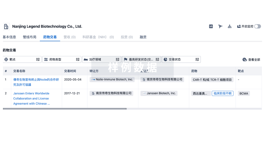
100 项与 GE Medical Systems Israel Ltd. 相关的药物交易
登录后查看更多信息
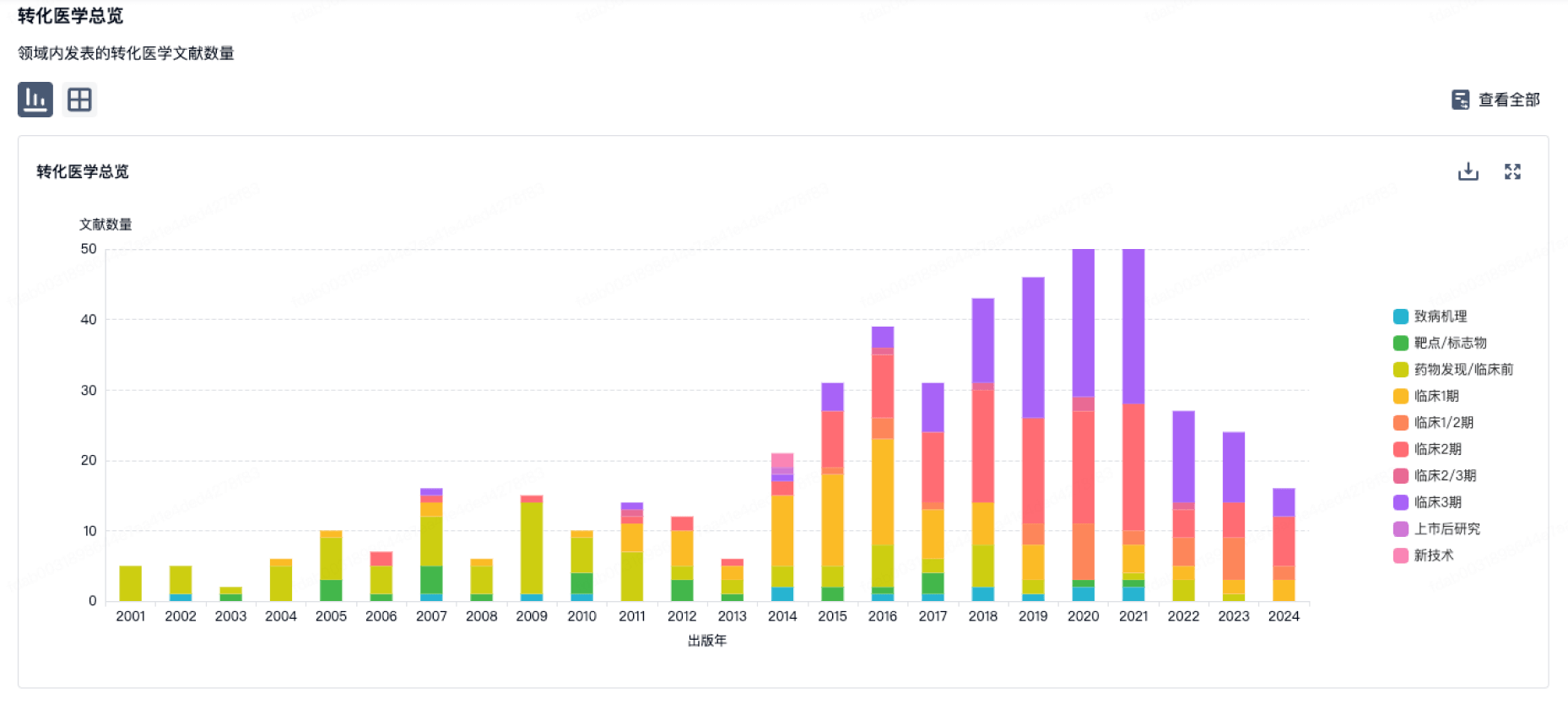
100 项与 GE Medical Systems Israel Ltd. 相关的转化医学
登录后查看更多信息
组织架构
使用我们的机构树数据加速您的研究。
登录
或

管线布局
2025年06月15日管线快照
无数据报导
登录后保持更新
药物交易
使用我们的药物交易数据加速您的研究。
登录
或
转化医学
使用我们的转化医学数据加速您的研究。
登录
或
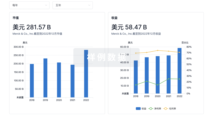
营收
使用 Synapse 探索超过 36 万个组织的财务状况。
登录
或
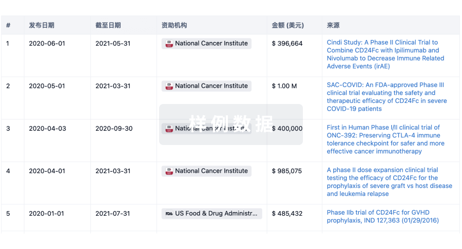
科研基金(NIH)
访问超过 200 万项资助和基金信息,以提升您的研究之旅。
登录
或
投资
深入了解从初创企业到成熟企业的最新公司投资动态。
登录
或
融资
发掘融资趋势以验证和推进您的投资机会。
登录
或
Eureka LS:
全新生物医药AI Agent 覆盖科研全链路,让突破性发现快人一步
立即开始免费试用!
智慧芽新药情报库是智慧芽专为生命科学人士构建的基于AI的创新药情报平台,助您全方位提升您的研发与决策效率。
立即开始数据试用!
智慧芽新药库数据也通过智慧芽数据服务平台,以API或者数据包形式对外开放,助您更加充分利用智慧芽新药情报信息。
生物序列数据库
生物药研发创新
免费使用
化学结构数据库
小分子化药研发创新
免费使用