预约演示
更新于:2025-09-06
Lysosomal Storage Disease Therapeutics(Orphazyme Aps)
更新于:2025-09-06
概要
基本信息
在研机构- |
权益机构- |
最高研发阶段无进展临床前 |
首次获批日期- |
最高研发阶段(中国)- |
特殊审评- |

关联
100 项与 Lysosomal Storage Disease Therapeutics(Orphazyme Aps) 相关的临床结果
登录后查看更多信息
100 项与 Lysosomal Storage Disease Therapeutics(Orphazyme Aps) 相关的转化医学
登录后查看更多信息
100 项与 Lysosomal Storage Disease Therapeutics(Orphazyme Aps) 相关的专利(医药)
登录后查看更多信息
5
项与 Lysosomal Storage Disease Therapeutics(Orphazyme Aps) 相关的文献(医药)2007-05-01·Reproductive sciences (Thousand Oaks, Calif.)3区 · 医学
Activation of Toll-like Receptors 2 or 3 and Preterm Delivery in the Mouse
3区 · 医学
Article
作者: Vladimir Ilievski ; Shi-Jiang Lu ; Emmet Hirsch
The objective of this study is to test whether the activation of toll-like receptors (TLRs) 2 and 3 (innate immune receptors for gram-positive and viral pathogens, respectively) can induce preterm delivery. One uterine horn of preterm pregnant CD-1 mice at approximately 75% of gestation was injected with TLR-2 ligands (lipoteichoic acid [LTA] or peptidoglycan [PGN]) or the TLR-3 ligand polyinosinic:cytidylic acid (poly[I:C]). Preterm delivery was recorded. In a separate group of mice, tissue mRNAs were quantified by reverse transcriptase polymerase chain reaction 5 hours after treatment with PGN or poly(I:C). Intrauterine PGN and LTA induced preterm delivery, reaching 100% at maximal doses. Intraperitoneal PGN also induced preterm delivery but at lower rates (maximum = 55%). Intrauterine poly(I:C) induced preterm birth in up to 31% of mice. Poly(I:C) induced uterine interferon beta and chemokine (C-C motif) ligand 5 (CCL5, also known as RANTES) but not interleukin 1beta, tumor necrosis factor, or lipopolysaccharide-induced CXC chemokine. PGN did not alter these mRNAs when compared with saline. Neither treatment induced gene expression in fetal membranes. Activation of either TLR-2 or -3 can induce preterm delivery in the mouse. Activation of TLR-3 with poly(I:C) induces interferon beta and the chemokine CCL5 in uterine tissues but not in fetal membranes.
2006-01-15·Journal of immunology (Baltimore, Md. : 1950)2区 · 医学
Anaphylactic Release of Mucosal Mast Cell Granule Proteases: Role of Serpins in the Differential Clearance of Mouse Mast Cell Proteases-1 and -2
2区 · 医学
Article
作者: Knight, Pamela A. ; Wright, Steven H. ; Pemberton, Alan D. ; Miller, Hugh R. P.
Abstract:
The granule-derived mouse mast cell proteases-1 and -2 (mMCP-1 and -2) colocalize in similar quantities in mucosal mast cells but micrograms of mMCP-1 compared with nanograms of mMCP-2 are detected in peripheral blood during intestinal nematode infection. This differential systemic response was investigated both in vitro and in vivo. Bone marrow-derived mucosal mast cell homologs released similar quantities of mMCP-1 and-2 concomitantly with β-hexosaminidase in response to calcium ionophore (∼60% release) or IgE/DNP (25% release). In contrast, serum from mice sensitized by infection with Nippostrongylus brasiliensis 10 days earlier contained >1500-fold more mMCP-1 (10,130 ± 1,609 ng/ml) than mMCP-2 (6.4 ± 1 ng/ml), but, in gut lumen, the difference was ∼8-fold. After OVA sensitization, >600-fold more mMCP-1 (7,861 ± 2,209 ng/ml) than mMCP-2 (12.8 ± 4.7 ng/ml) was present in blood 1 h after challenge, but, in gut lumen, there were relatively comparable levels of mMCP-1 and -2. To estimate the rates of systemic accumulation and clearance, 10 μg of mMCP-1 or -2 was injected i.p. Plasma levels of injected mMCP-2 peaked (1%) at 15 min then declined, whereas levels of mMCP-1 were maximal (∼25%) at 3 h. Inactivation of mMCP-1 with PMSF before injection resulted in mMCP-2-like kinetics, but inhibition of mMCP-1 by serum gave kinetics similar to that of native mMCP-1. mMCP-1 isolated from serum is complexed with serpins and we conclude that both the accumulation and the longevity of mMCP-1 in blood is due to complex formation, protecting it from a pathway that rapidly clears mMCP-2, which is unable to form complexes with serpins.
2004-01-01·American journal of physiology. Gastrointestinal and liver physiology3区 · 医学
Anti-inflammatory effect of two isoforms of COX inH. pylori-induced gastritis in mice: possible involvement of PGE2
3区 · 医学
Article
作者: Higuchi, Kazuhide ; Watanabe, Toshio ; Tanigawa, Tetsuya ; Fujiwara, Yasuhiro ; Sasaki, Eiji ; Matsumoto, Takayuki ; Hamaguchi, Masaki ; Tominaga, Kazunari ; Arakawa, Tetsuo ; Oshitani, Nobuhide
Neutrophil infiltration mediated by TNF-α is associated with various types of gastric injury, whereas PGs play a crucial role in gastric defense. We examined roles of two isoforms of cyclooxygenase (COX) and PGE2in Helicobacter pylori-induced gastritis in mice. Mice infected with H. pylori were given selective COX-1 inhibitor SC-560 (10 mg/kg), selective COX-2 inhibitor NS-398 (10 mg/kg), or nonselective COX inhibitor indomethacin (2 mg/kg) with or without 16,16-dimethyl PGE2for 1 wk. H. pylori infection increased levels of mRNA for COX-1 and -2 in gastric tissue by 1.2-fold and 3.3-fold, respectively, accompanied by a significant increase in PGE2production by gastric tissue. H. pylori infection significantly elevated MPO activity, a marker of neutrophil infiltration, and epithelial cell apoptosis in the stomach. SC-560 augmented MPO activity and epithelial cell apoptosis with associated reduction in PGE2production, whereas NS-398 had the same effects without affecting PGE2production. Inhibition of both COX-1 and -2 by indomethacin or concurrent treatment with SC-560 and NS-398 resulted in a stronger increase in MPO activity and apoptosis than inhibition of either COX-1 or -2 alone. H. pylori infection elevated TNF-α mRNA expression in the stomach, which was further increased by indomethacin. Effects of COX inhibitors on neutrophil infiltration, apoptosis, and TNF-α expression in H. pylori-infected mice were abolished by exogenous 16,16-dimethyl PGE2. In conclusion, PGE2derived from either COX-1 or -2 is involved in regulation of gastric mucosal inflammation and contributes to maintenance of mucosal integrity during H. pylori infection via inhibition of TNF-α expression.
100 项与 Lysosomal Storage Disease Therapeutics(Orphazyme Aps) 相关的药物交易
登录后查看更多信息
研发状态
10 条进展最快的记录, 后查看更多信息
登录
| 适应症 | 最高研发状态 | 国家/地区 | 公司 | 日期 |
|---|---|---|---|---|
| 溶酶体贮积病 | 临床前 | 丹麦 | - |
登录后查看更多信息
临床结果
临床结果
适应症
分期
评价
查看全部结果
| 研究 | 分期 | 人群特征 | 评价人数 | 分组 | 结果 | 评价 | 发布日期 |
|---|
No Data | |||||||
登录后查看更多信息
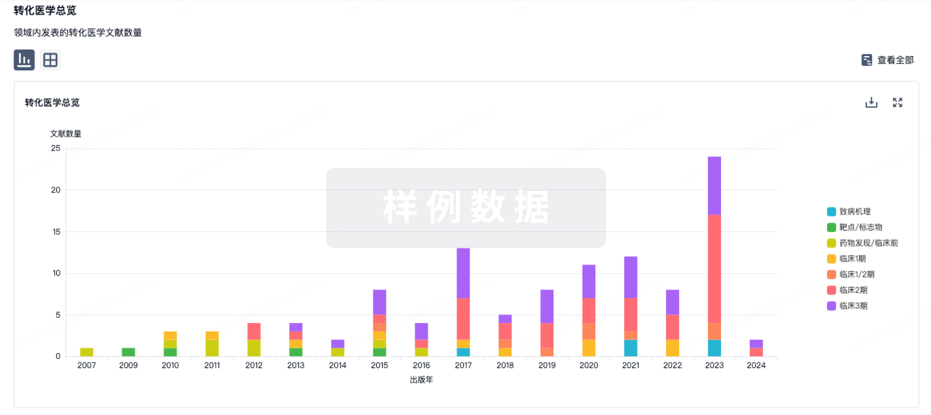
转化医学
使用我们的转化医学数据加速您的研究。
登录
或
药物交易
使用我们的药物交易数据加速您的研究。
登录
或
核心专利
使用我们的核心专利数据促进您的研究。
登录
或
临床分析
紧跟全球注册中心的最新临床试验。
登录
或
批准
利用最新的监管批准信息加速您的研究。
登录
或
生物类似药
生物类似药在不同国家/地区的竞争态势。请注意临床1/2期并入临床2期,临床2/3期并入临床3期
登录
或
特殊审评
只需点击几下即可了解关键药物信息。
登录
或
生物医药百科问答
全新生物医药AI Agent 覆盖科研全链路,让突破性发现快人一步
立即开始免费试用!
智慧芽新药情报库是智慧芽专为生命科学人士构建的基于AI的创新药情报平台,助您全方位提升您的研发与决策效率。
立即开始数据试用!
智慧芽新药库数据也通过智慧芽数据服务平台,以API或者数据包形式对外开放,助您更加充分利用智慧芽新药情报信息。
生物序列数据库
生物药研发创新
免费使用
化学结构数据库
小分子化药研发创新
免费使用